 |
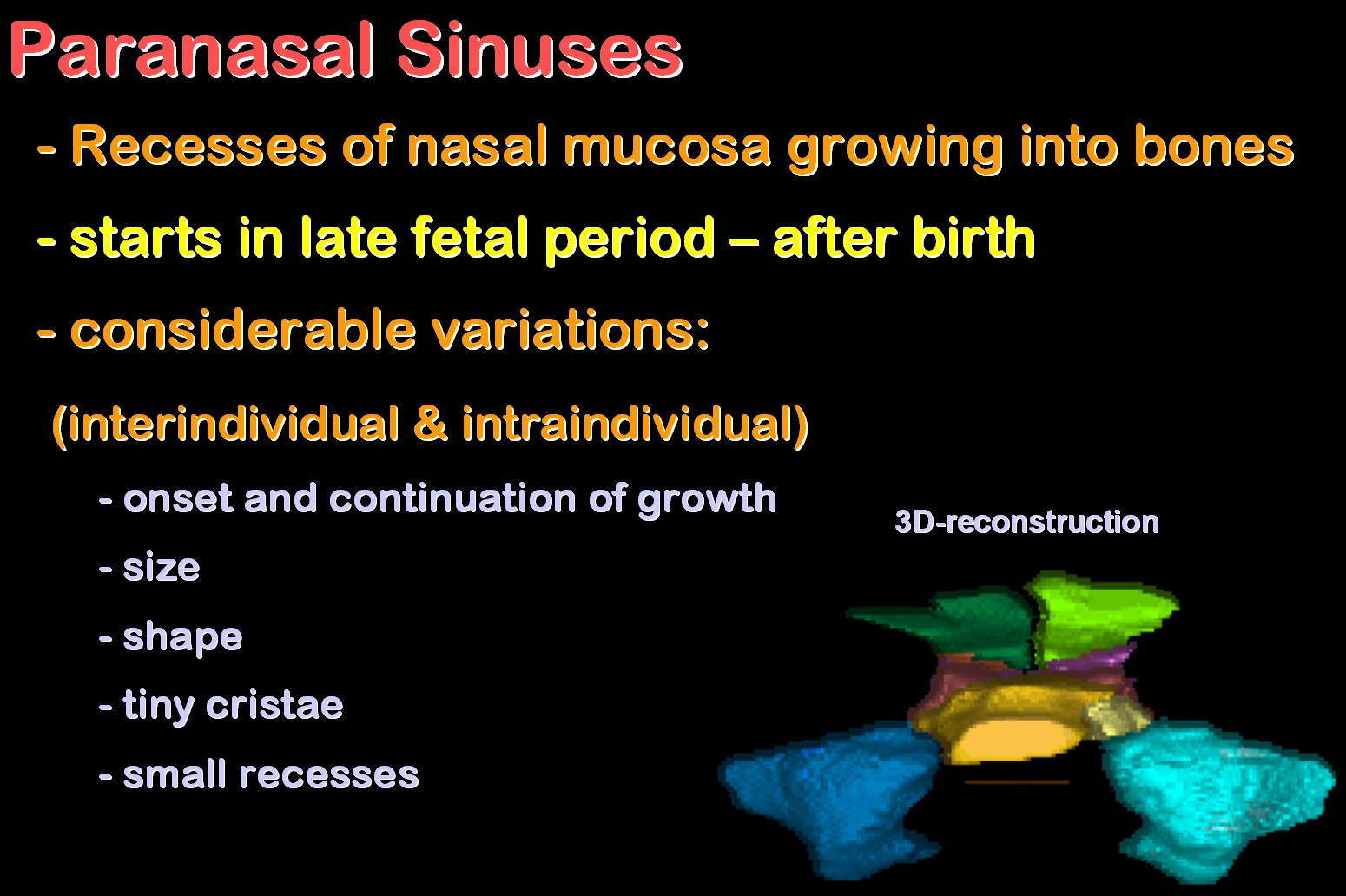
The paranasal sinuses (PNS)
are recesses of nasal mucosa growing into greater bones lateral or superior
to the nasal cavity. This process starts in late fetal period or after
birth. There are considerable variations both, interindividual and intraindividual
concerning the onset and continuation of growth, the size and the shape
of these air sacs which have variable cristae in their lumen and may form
small recesses.
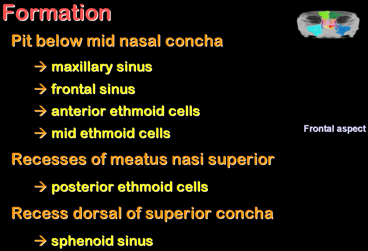
During development, a small pit below mid nasal concha gives raise to the maxillary and frontal sinus as well as to the anterior and mid ethmoid cells. A recesses of the upper nasal duct is the origin of the posterior ethmoid cells, and a recess dorsal of superior concha grows into the sphenoid bone to form its sinus. Click here to see the animated 3D reconstructions |
 |
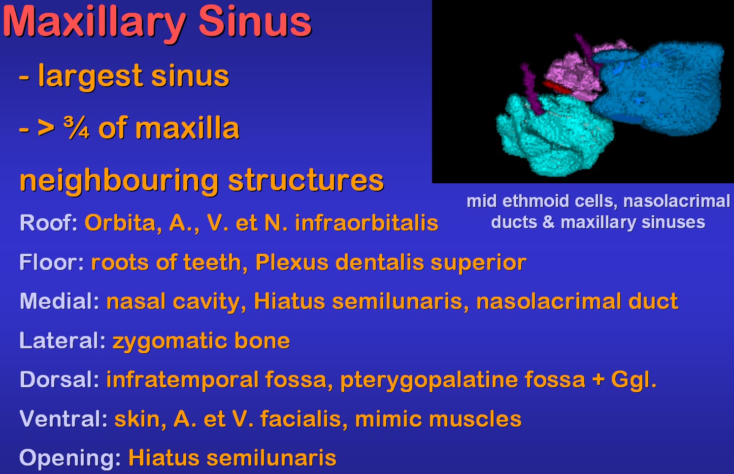 |
The maxillary sinus, which fills
over three quarters of the bone, is the largest PNS. Important neighbouring
structures are shown on the left, while on the right, blood vessels and
nerves of this sinus are listed.
Click here to see the animated 3D reconstruction |
 |
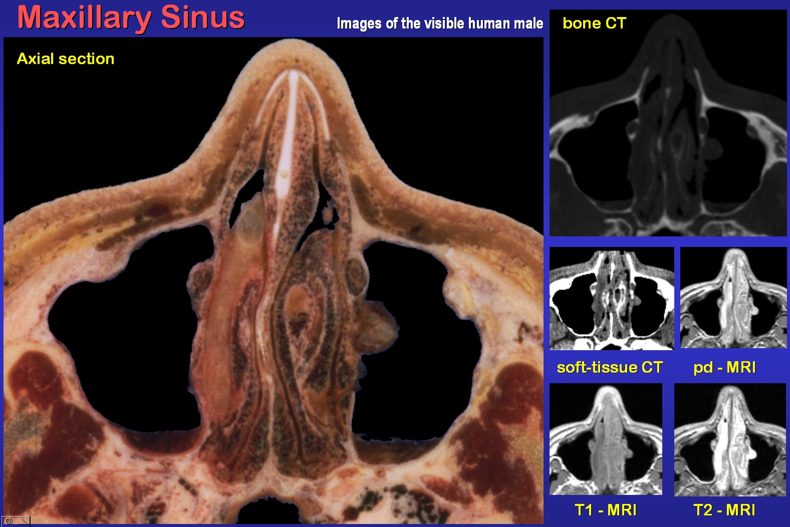 |
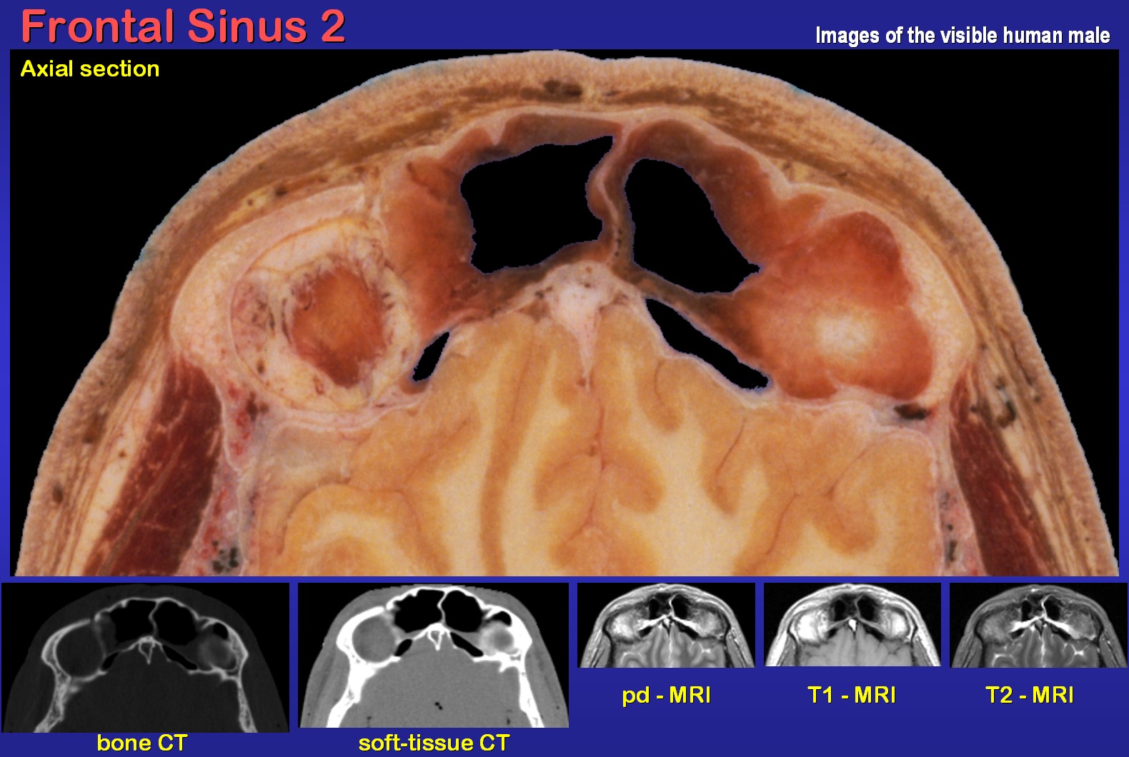
An axial section through the
maxillary sinus and corresponding radiological images of the visible human
(vh) male are demonstrated on the left.
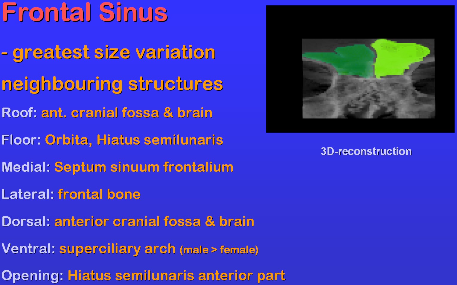
The frontal sinus has the greatest variation in size (agenesia to lumina far over 10 ml). Its most important neighbouring structures are noted on the right. Click here to see the animated 3D reconstruction |
 |
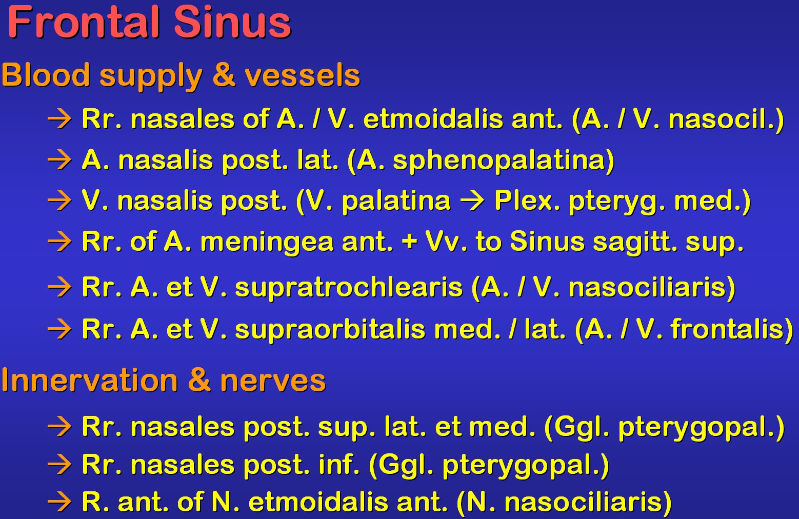 |
There are some small vessels
and nerve branches for the frontal sinus. Their origin is shown on the
left.
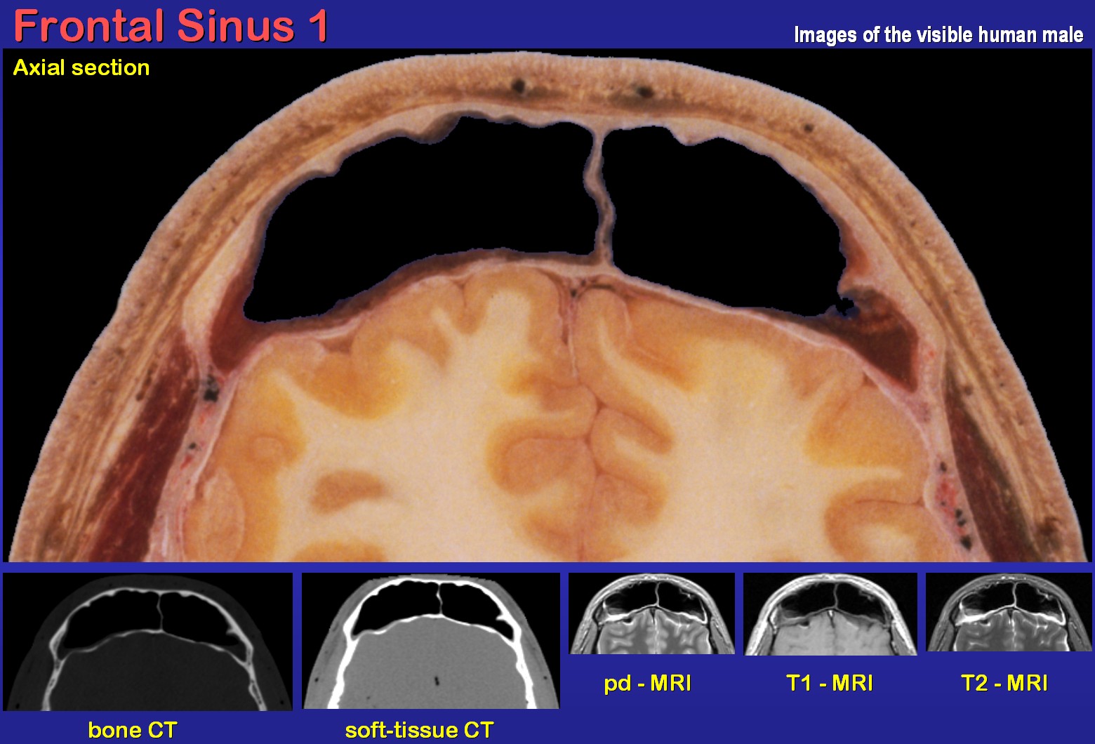
As visible on the right, the vh male has quite large frontal sinuses. Note that the septum between them is not quite in the median plane. The posterior wall of the frontal sinuses is only a thin bony lamella in close vicinity to the brain. |
 |
 |
The images on the left show
the same frontal sinuses further down towards the natural opening. The
left orbita is opened and the levator palpebrae muscle is cut. The right
orbital roof is the floor of the frontal sinus.
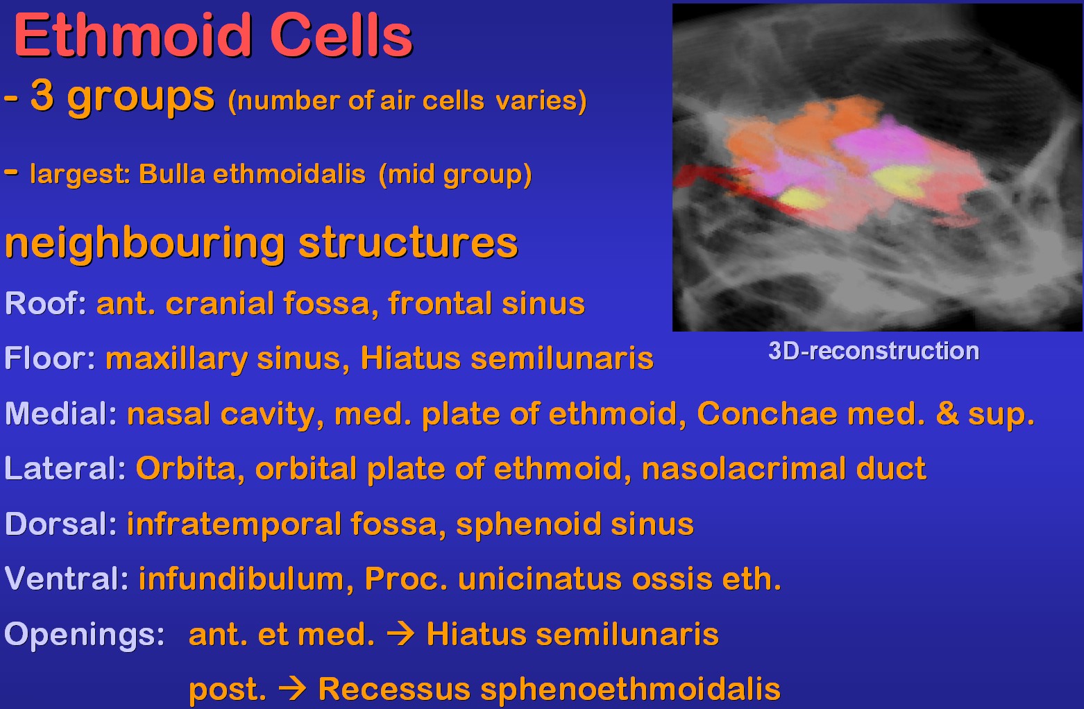
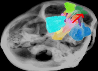
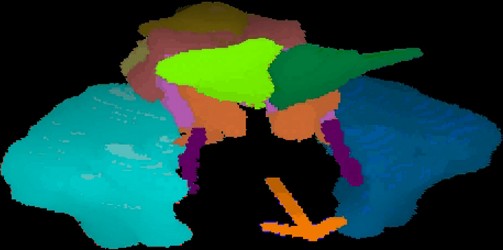
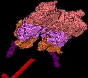
The 3D-reconstruction on the right shows the anterior ethmoid cells in orange, the mid ones in magenta and the posterior ones in purple. On each site the ethmoid bulla, which is the largest mid-ethmoid cell, is visible in yellow. Note the neighbouring structures and that the natural apertures of the anterior and mid ethmoid cells drain into the semilunar hiatus whereas the posterior ones open into the sphenoethmoid recess. Click here to see the animated 3D reconstruction |
 |
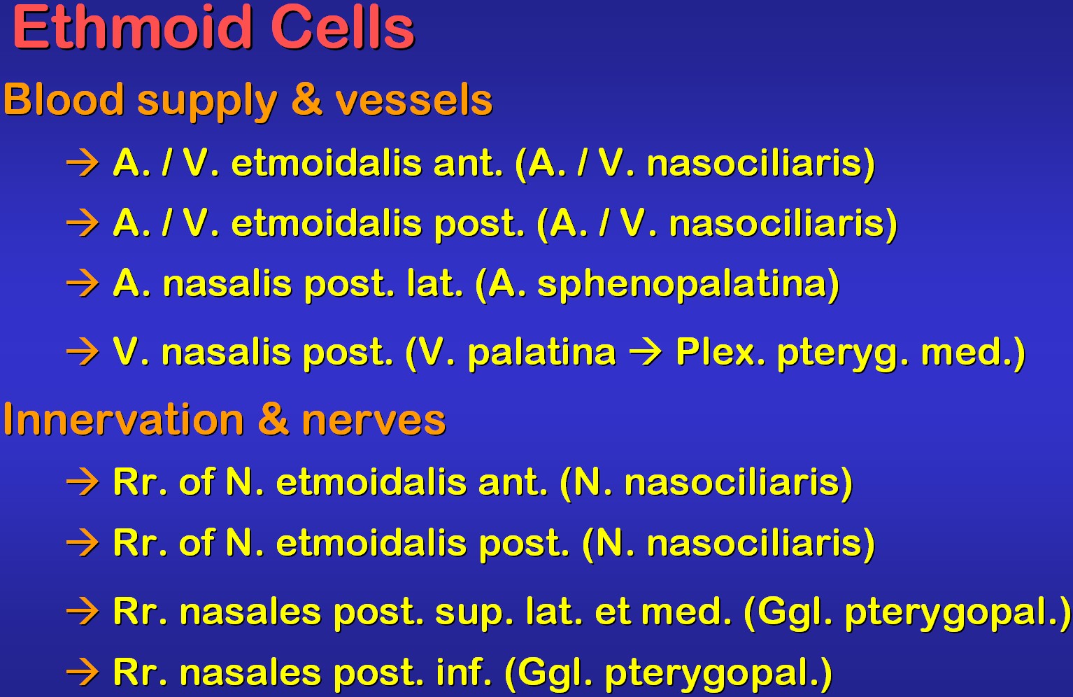 |
The vessels and nerves of the
ethmoid cells derive mostly from small branches of the nasociliar artery,
vein and nerve entering the sinuses via the anterior or posterior foramina
(left).
The posterior ethmoid cells directly border the sphenoid sinus. Note the carotid artery, pituitary and cavernous sinus dorsal to the sphenoid sinus and the rectus medialis muscle just lateral to the posterior ethmoid cells (right). |
 |
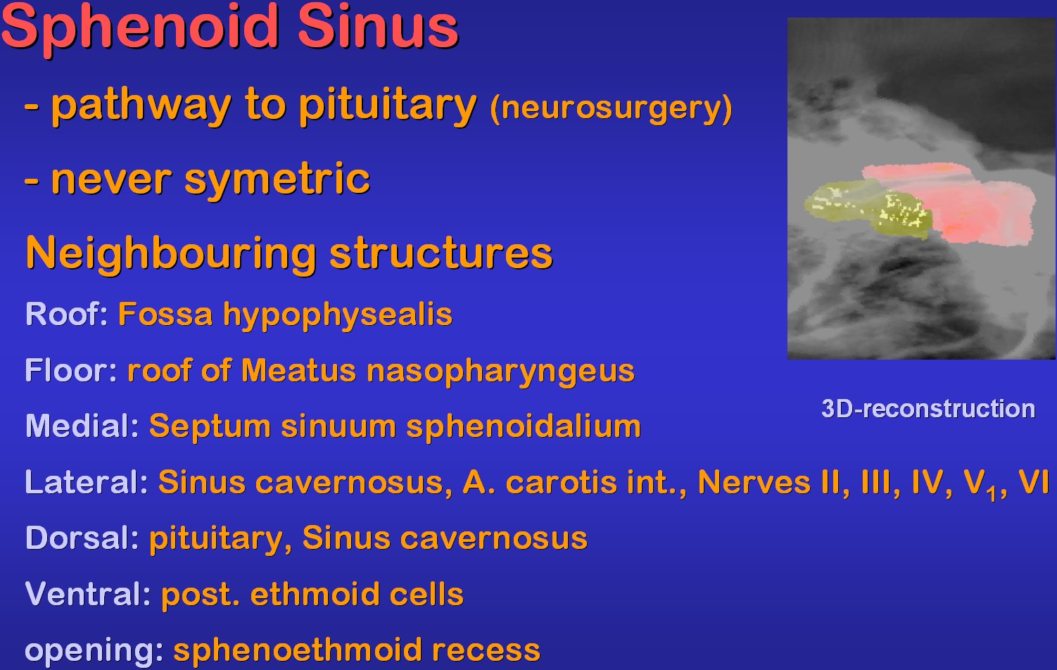 |
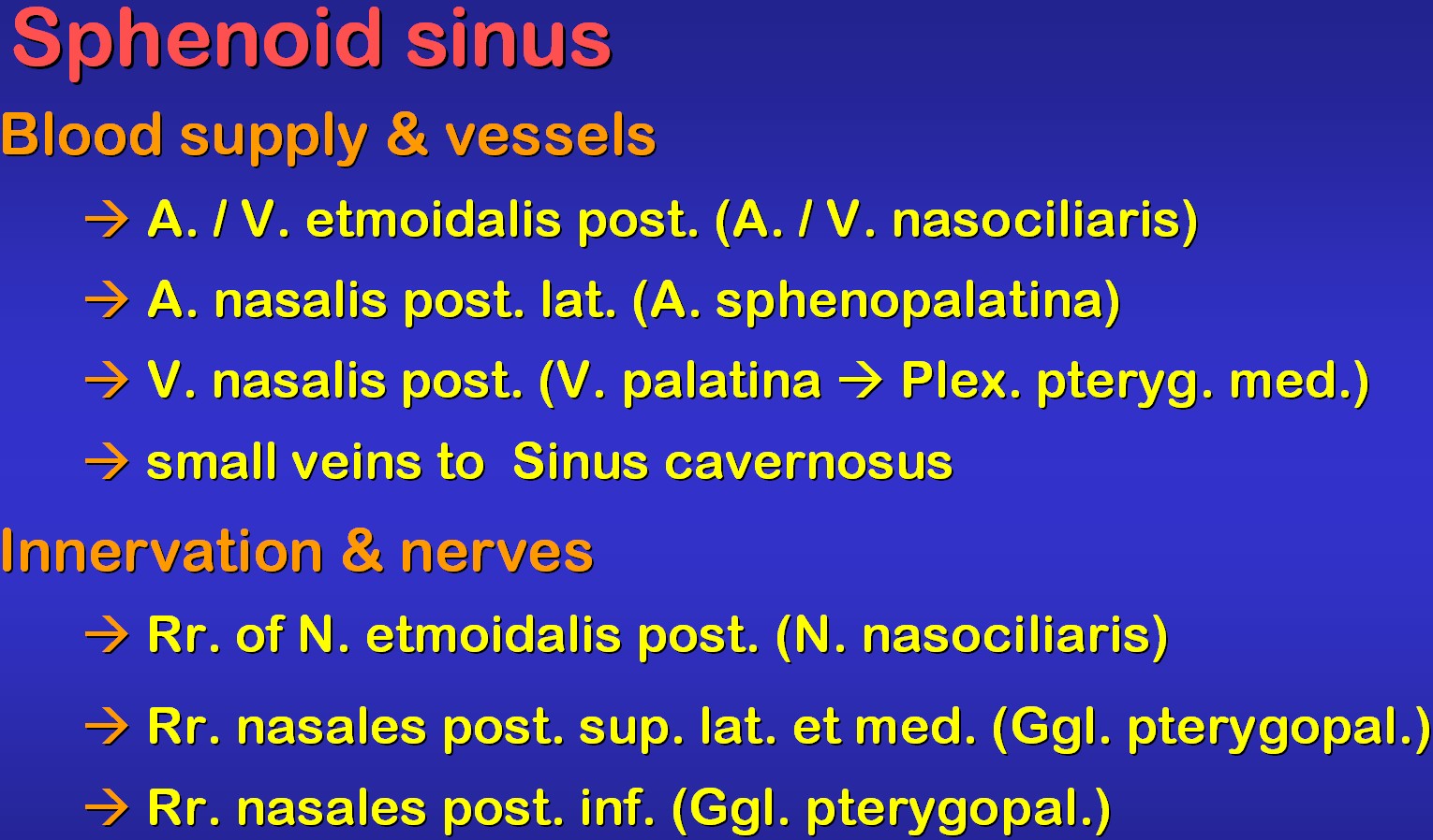
On the left a reconstruction
of both sphenoid sinuses of the vh female demonstrates the asymmetry of
the latter which are the most important pathway to the pituitary in neurosurgery.
There are many most impostant structures dorsolateral to the sinuses.
On the right structures for blood supply / drainage and innervation of the sinus are listed. Click here to see the animated 3D reconstruction |
 |
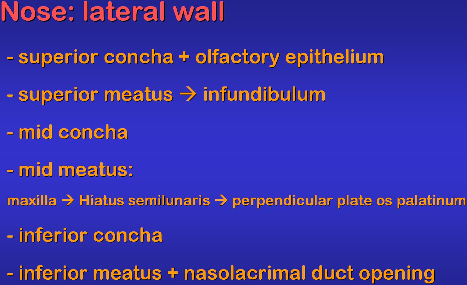 |
The lateral wall of the nose
is most important in PNS surgery. Some of its main structures are listed
on the left.
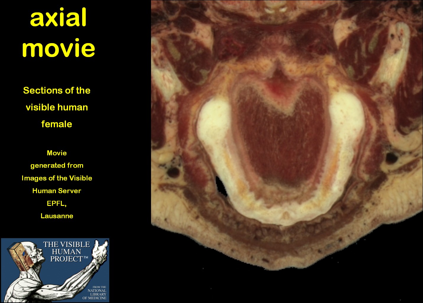
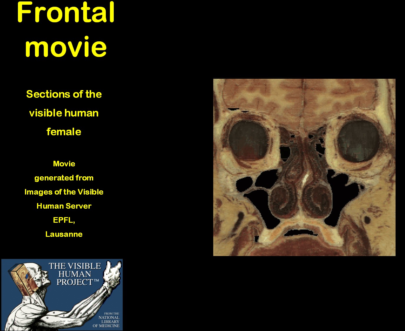
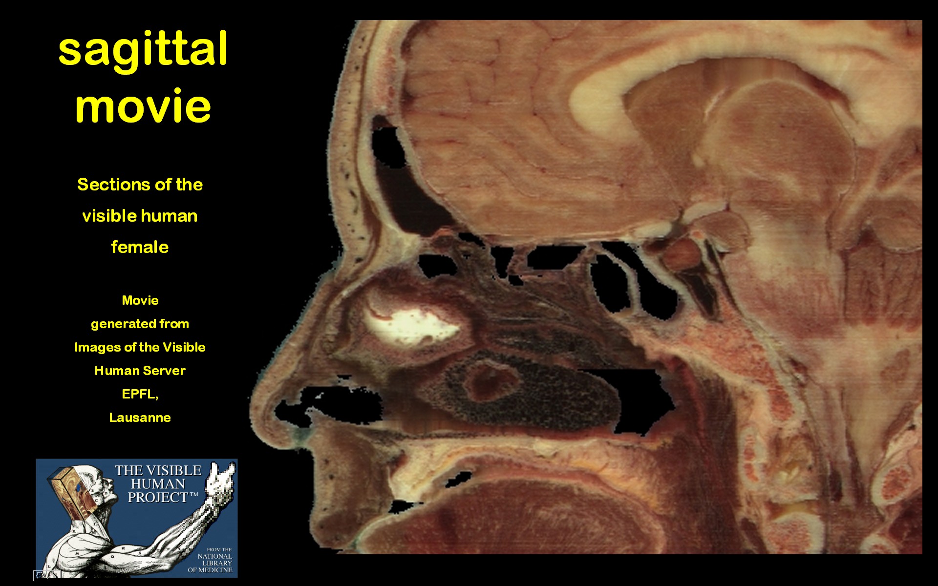
The vh female dataset with its sections cut at a distance of 0.33 mm is ideal for generation of motion pictures of original (axial) and computed (frontal/sagittal) true-colour sections in a good quality. In this way a three-dimensional picture of PNS topography can be obtained. Click here to see the movie |
 |
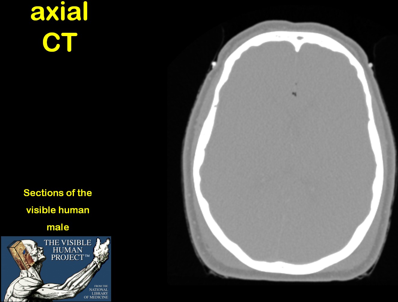 |
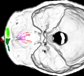
The images lead to motion pictures of the axial vh
female CT scans (left) and the segmented PNS of the 3D reconstructions
shown below (right).
Click here to see the motion pictures |
 |
 |
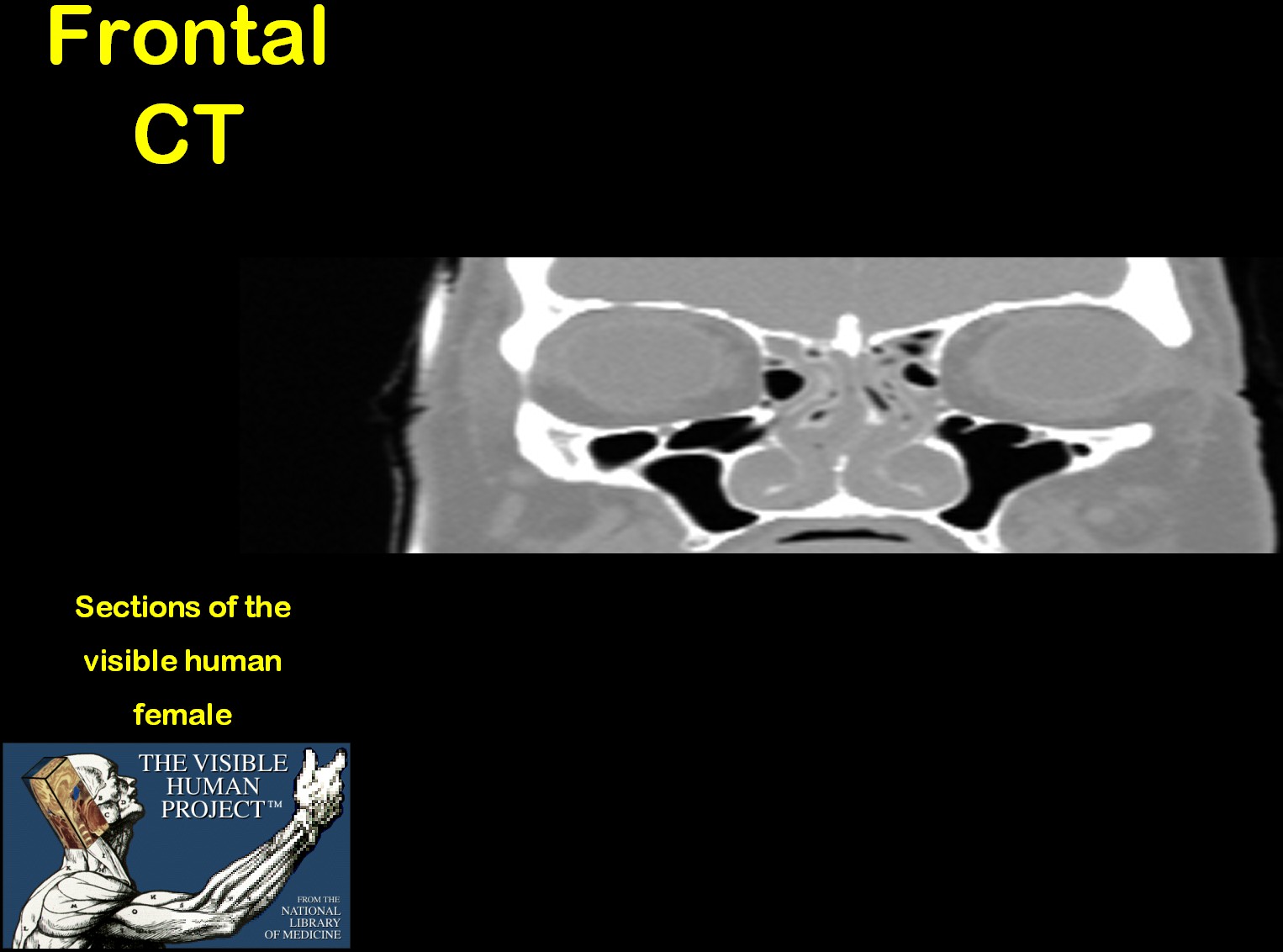
The computed frontal vh female sections and the frontal
CT sequences show e.g., that there are bony crests in the roof of the maxillary
sinus.
Click here to see the animations |
 |
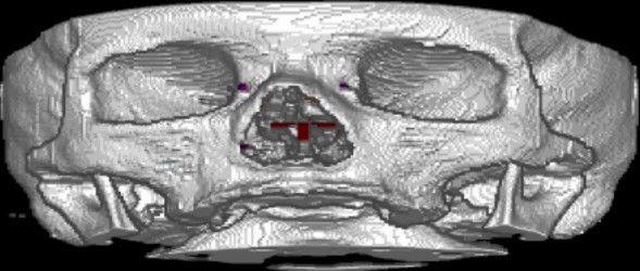 |
The images lead to motion pictures of the segmented
frontal vh female CT scans (left) and computed sagittal sections (right).
Click here to see the movies |
 |
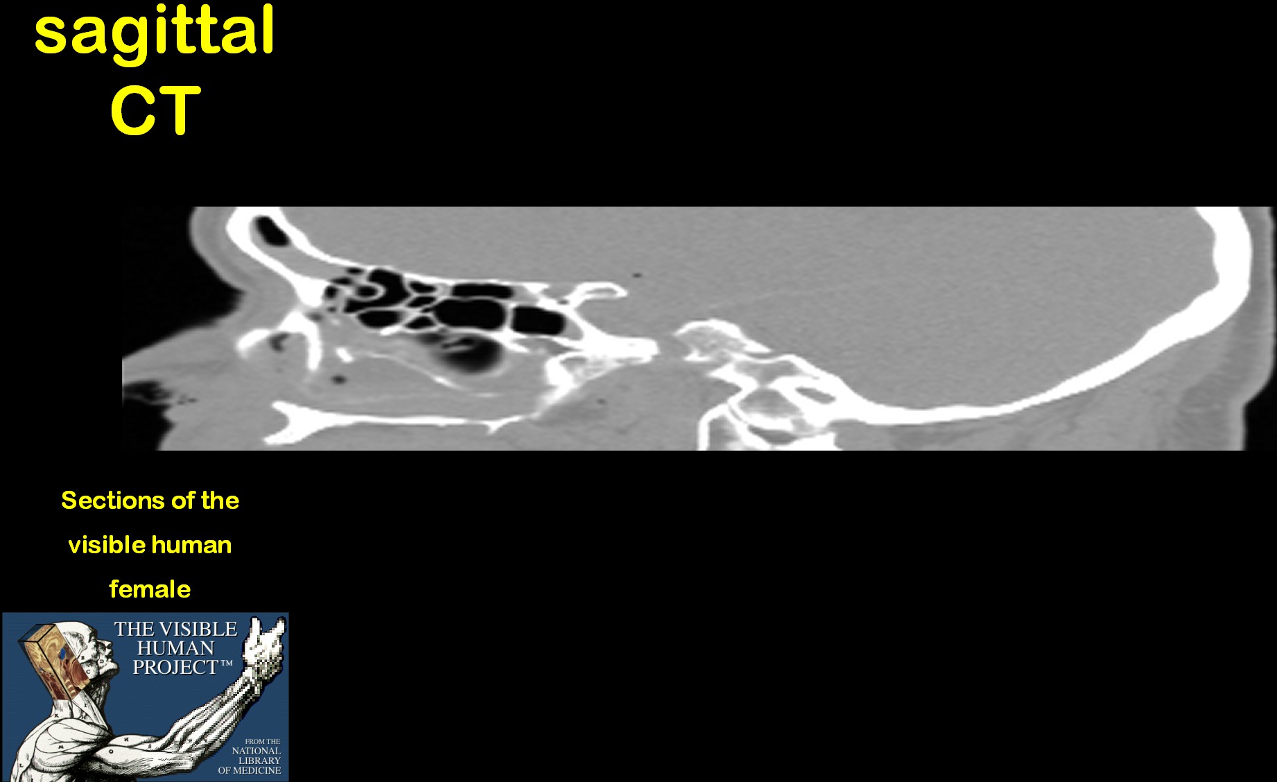 |
A movie of the sagittal vh female CT scans and one
of the segmented dataset is available.
Click here to see the motion pictures |
 |
 |
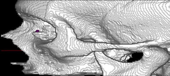
These images lead to movies navigating through the
reconstuction of the PNS of the vh female with transparent bones.
Click here to see the animated 3D reconstructions |
 |
 |
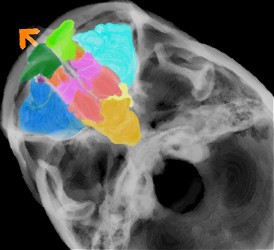
Whereas on the left, all PNS and the nasolacrimal
ducts (nld) are demonstrated in 3D, the movie linked to the right picture
focuses on the ethmoid cells and the nld.
Click here to see the animated 3D reconstructions |
 |