professional version of Dr. Jastrow's
Light- and Electron Microscopic Atlas
contains a few
movies of living
human sperm
cells

Link to the German
version of this page
|
professional version of Dr. Jastrow's Light- and Electron Microscopic Atlas |
|
This atlas also
contains a few movies of living human sperm cells |
|
Link to the German version of this page |
Short description:
This light- and electron microscopic atlas contains over 4,100 original
digital original images of very high quality and very
high resolution deriving from man, monkey or mammals. Comprehensive
texts provide detailed information on practically all
tissues,
organs, cell organelles & structures. Where possible one can explore
characteristics from light microscopic overview images via images in increasing
magnification to cell organelles which are only visible in electron microscopy
Plenty of pictures labelled in great detail. Interlinked texts and a vocabulary
which also contains background information on many biochemical
substances in English and German explain cellular structures in a generally
intelligible way and demonstrate interrelations as well as correlations
of structure and function. All images of this atlas can be quickly and
easily integrated in own presentations.
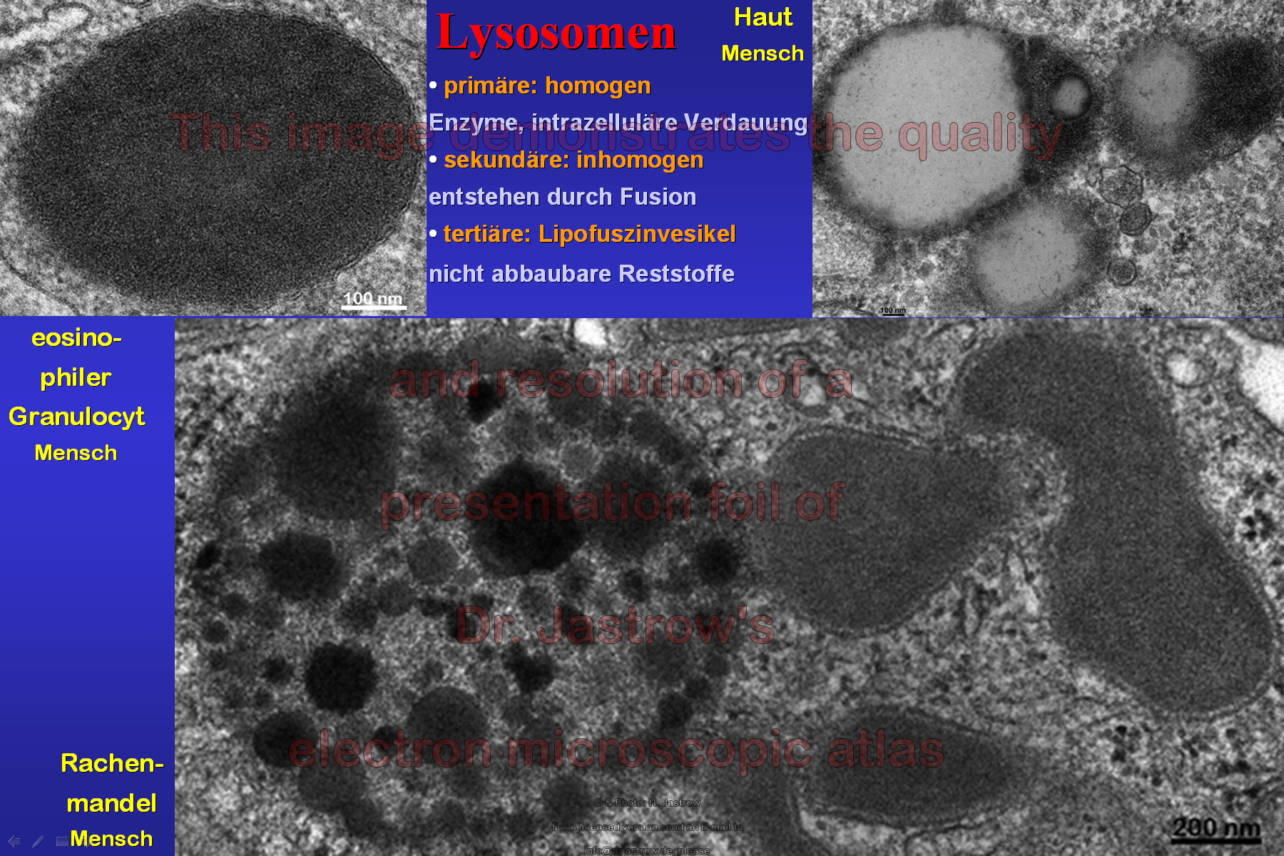
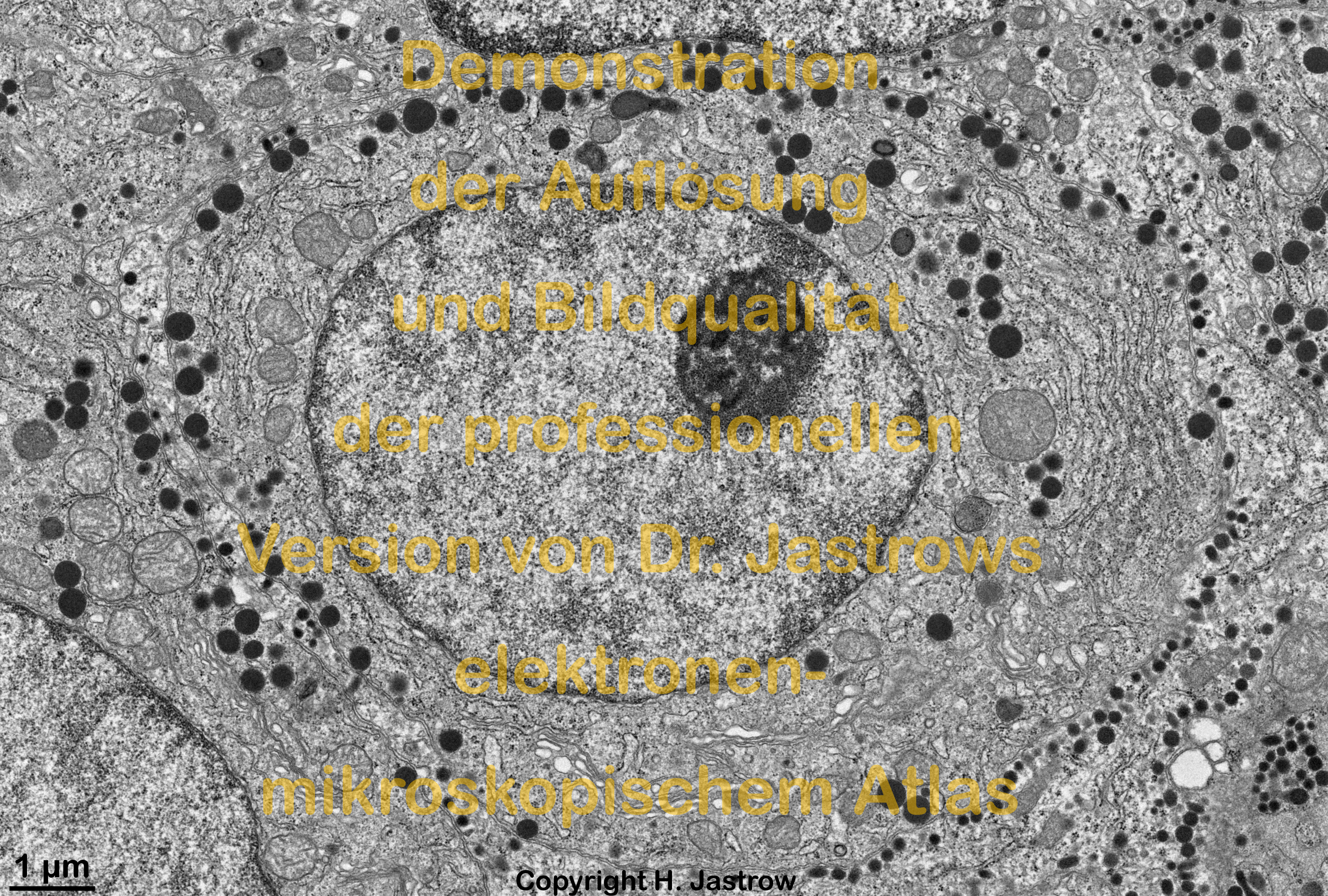
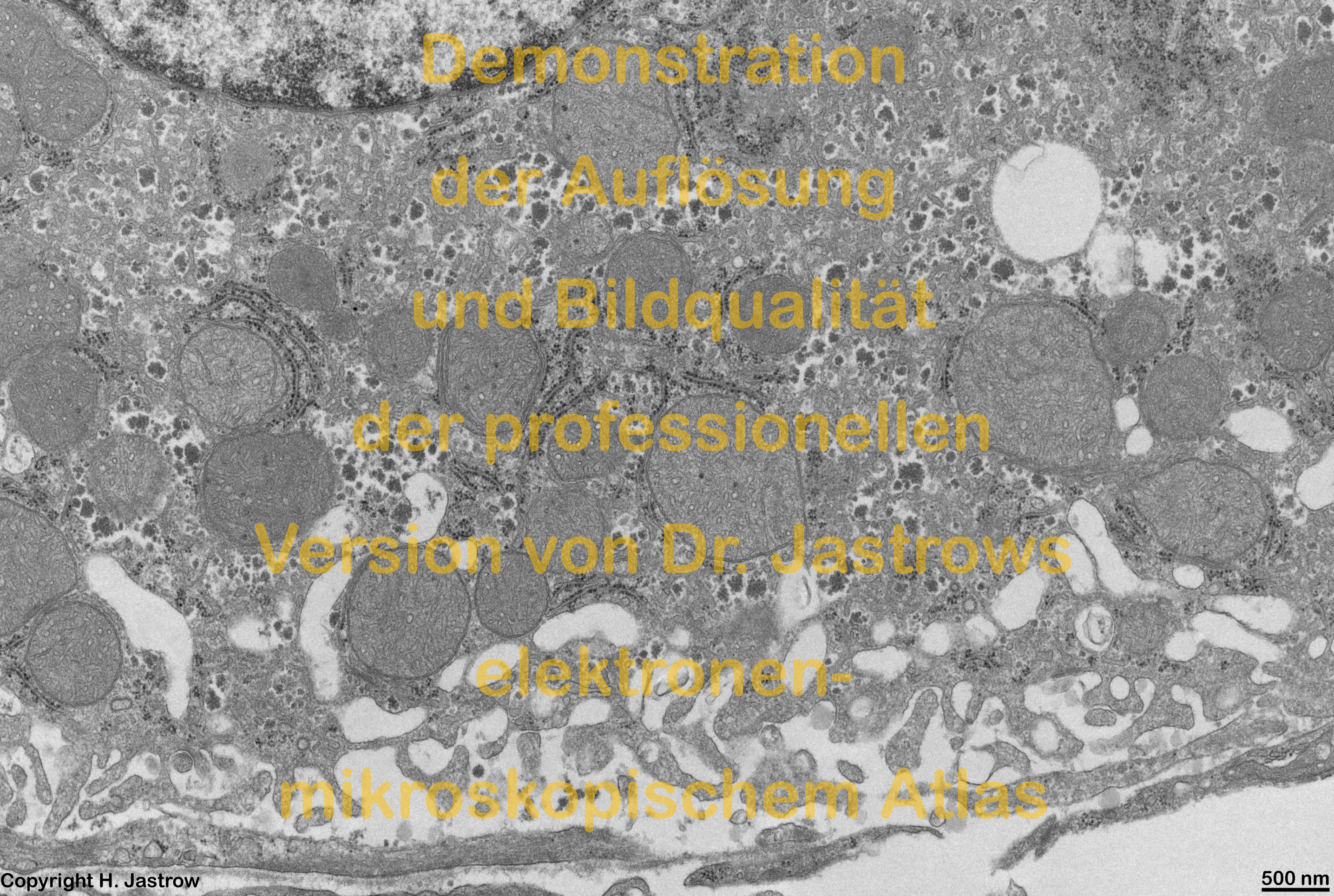
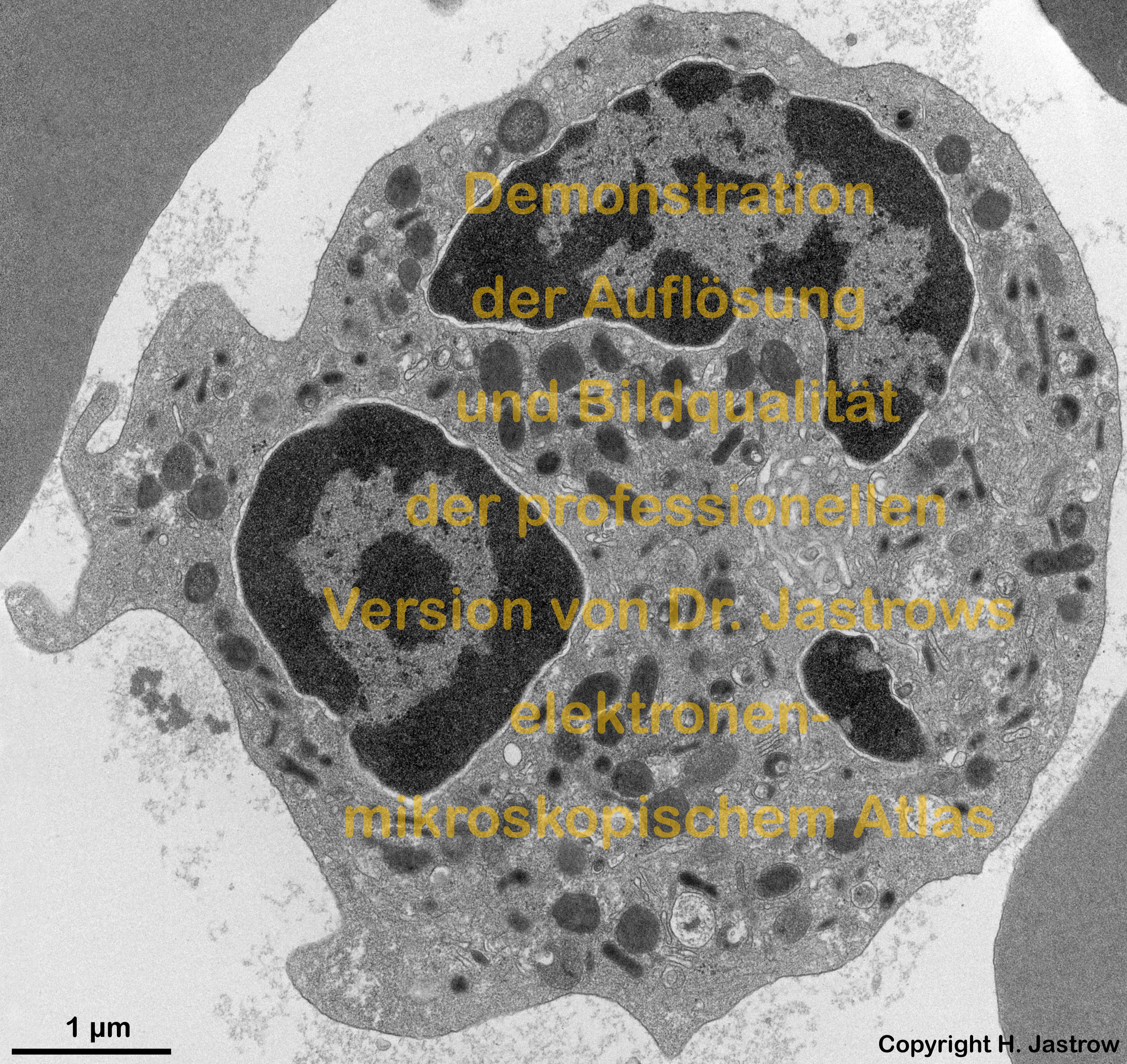
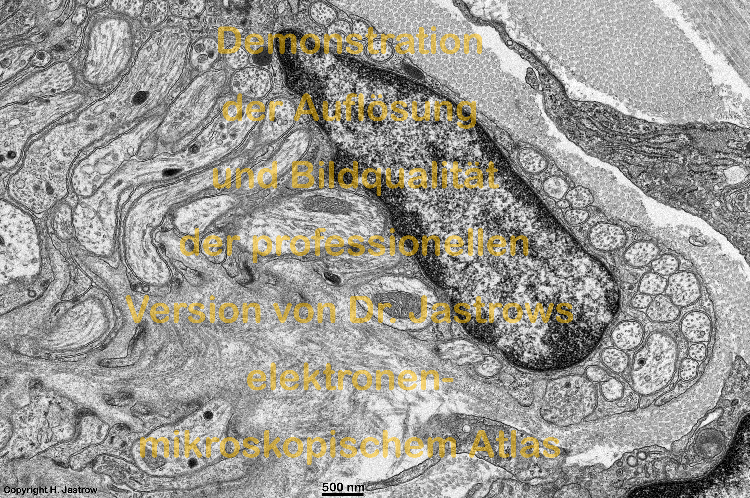
Demo pages in high resolution:
Selection of pages (screen snapshots) that demonstrate
the resolution and quality of the professional Version
(Clicking any miniaturized image calls up the original
sized image. The file size is given in MB)
| 1. Overview pages providing miniatures of original pictures (page from the www demo version with active links is loaded when lower text is clicked) | 2. Sample animation (click for high resolution) | ||||
 |
 |
 |
 |
 |
 |
|
Page of the www-version for comparison |
Page of the www-version for comparison |
Page of the www-version for comparison |
Page of the www-version for comparison |
Page of the www-version for comparison |
rod ribbon synapse 11.3 MB |
| 5. Labelled images (page with active links from the www demo version is loaded when the lower text is clicked) | 6. Lecture images (in German) | ||||||
 |
 |
 |
 |
 |
 |
 |
|
|
Seite der WWW-Version zum Vergleich |
Seite der WWW-Version zum Vergleich |
Seite der WWW-Version zum Vergleich |
Seite der WWW-Version zum Vergleich |
Seite der WWW-Version zum Vergleich |
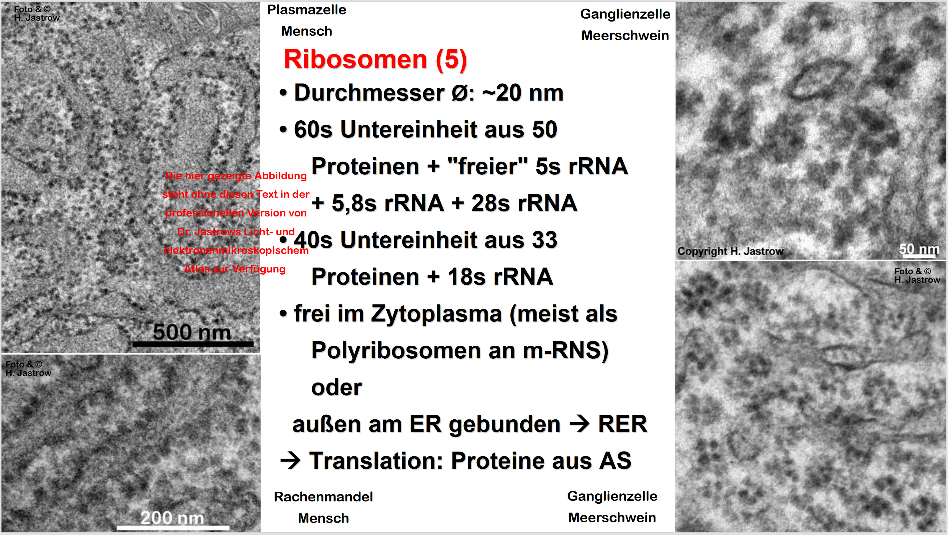
Zellstrukturen 1,9 MB - nicht im www verfügbar |
Ribosomen zeigt 1,4 MB - nicht im www verfügbar |
|
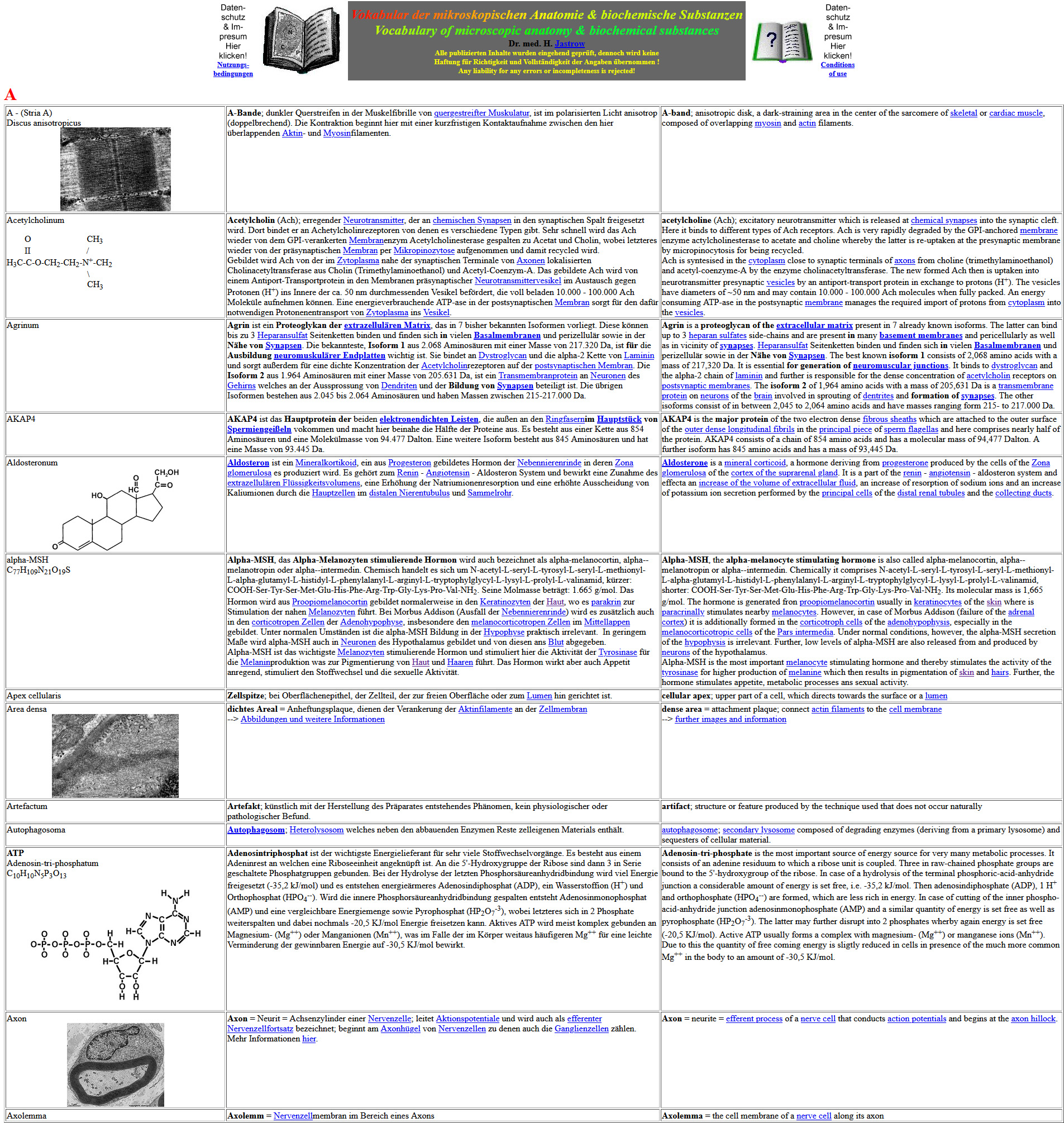
| 7. Page of the vocabulary
(only the initial part of the page is shown) |
8. examples of some new pages only present in the professional version (Please click on the thumbnails for higher resolution! Only the beginning of the pages with the overview of available images is shown) | ||||||
 |
 |
 |
 |
 |
 |
 |
|
|
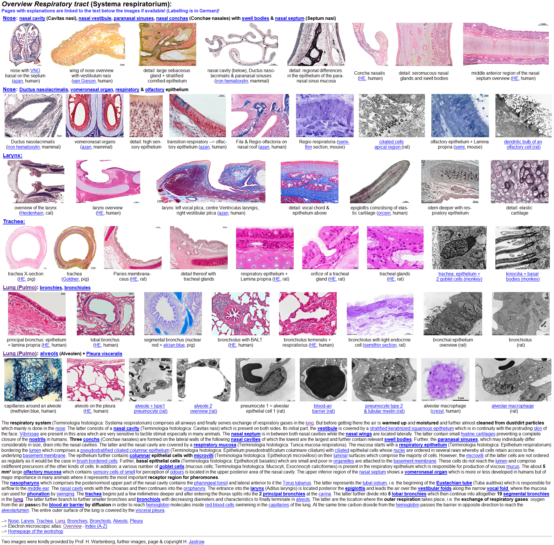
Seite der WWW-Version zum Vergleich |
Respiratory Tract* |
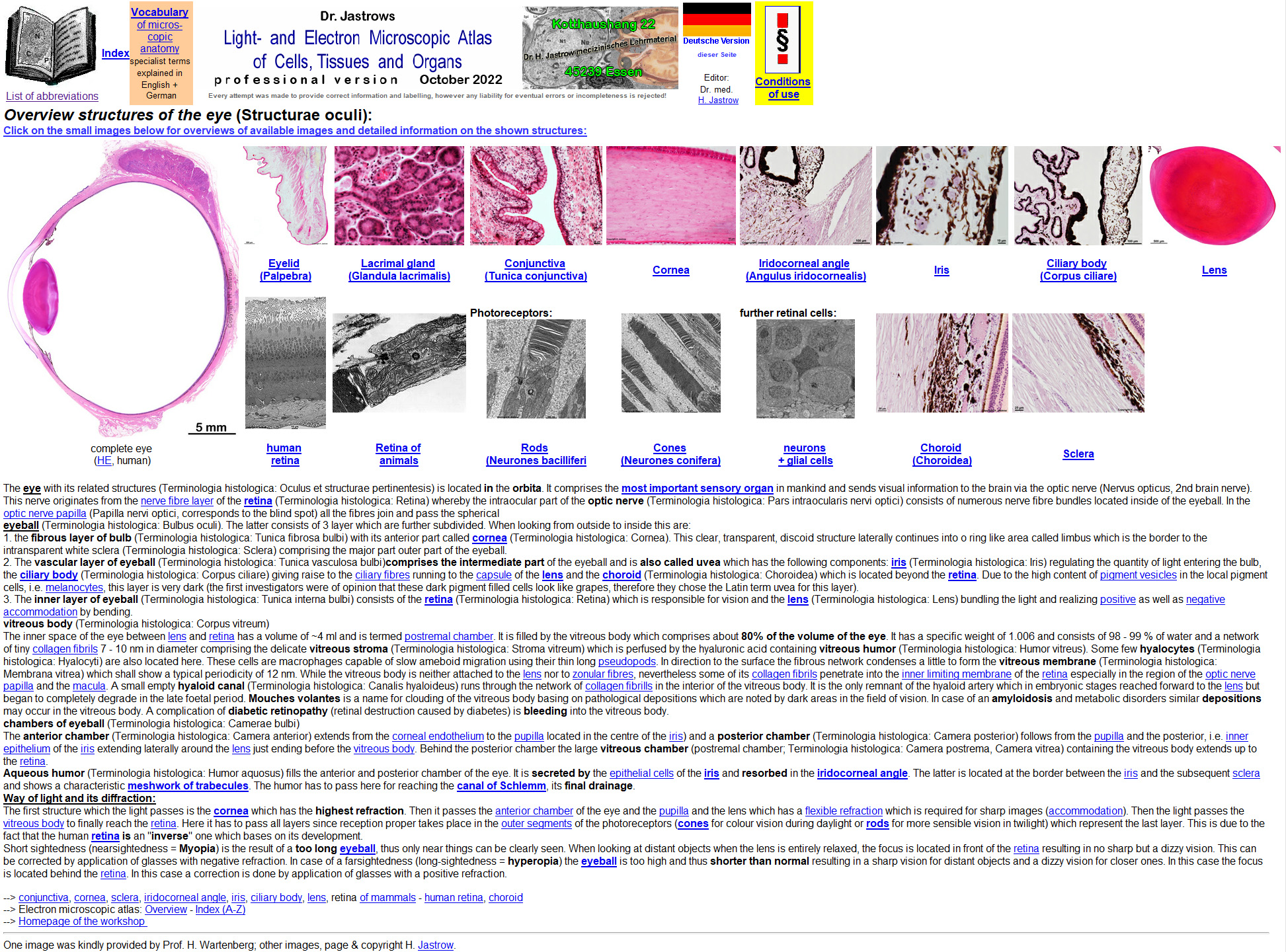
Eye and eye structures* |
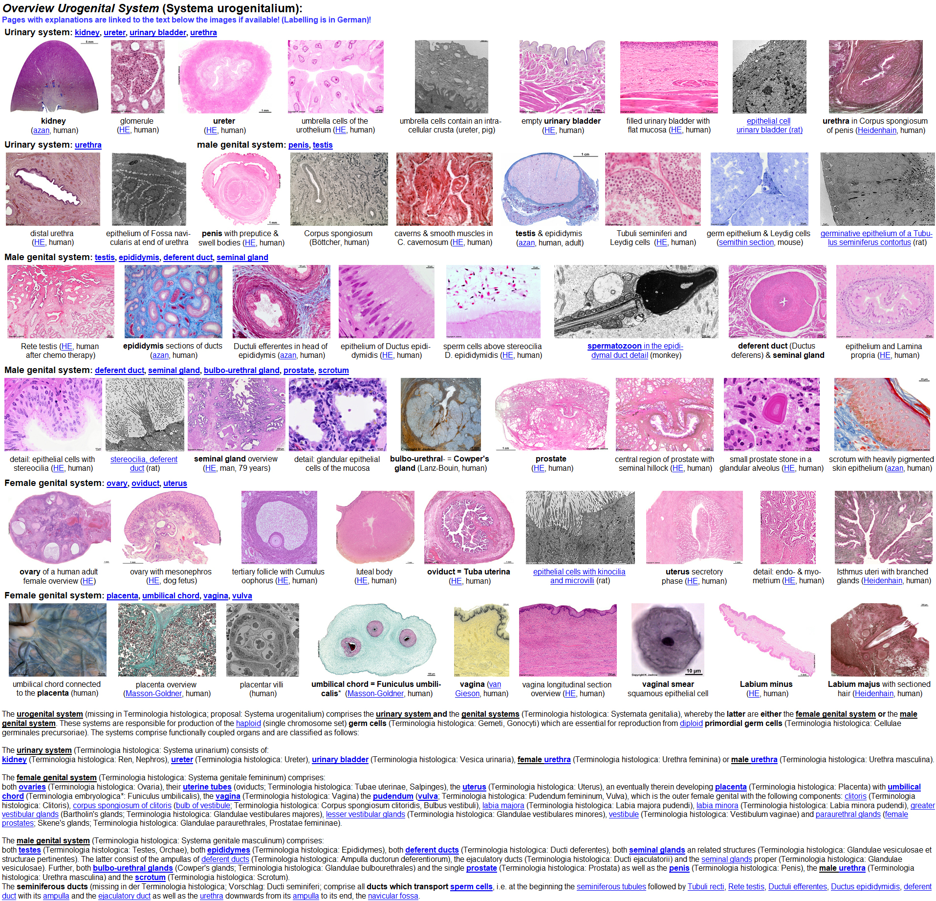
Urogenital tract* |
|
|
|
|
Detailed description:
Anatomical basic (and special) knowledge is of major impact in basic
and further education. It is an important basis for understanding medicine,
physiology, common and molecular cell biology, pathology, biochemistry,
pharmacology and further disciplines of the biomedical instruction and
research field. In this context a profound knowledge of cell and tissue
ultrastructures (structures which are too small to be visible in the light
microscope) is an excellent key to understand life processes and the reason
of many diseases. In this context original images of splendid quality and
resolution showing real ultrastructures are essential. However there are
few text books or other sources of images available which offer sufficiently
detailed electron-micrographs in a number that allows to get a general
overview.
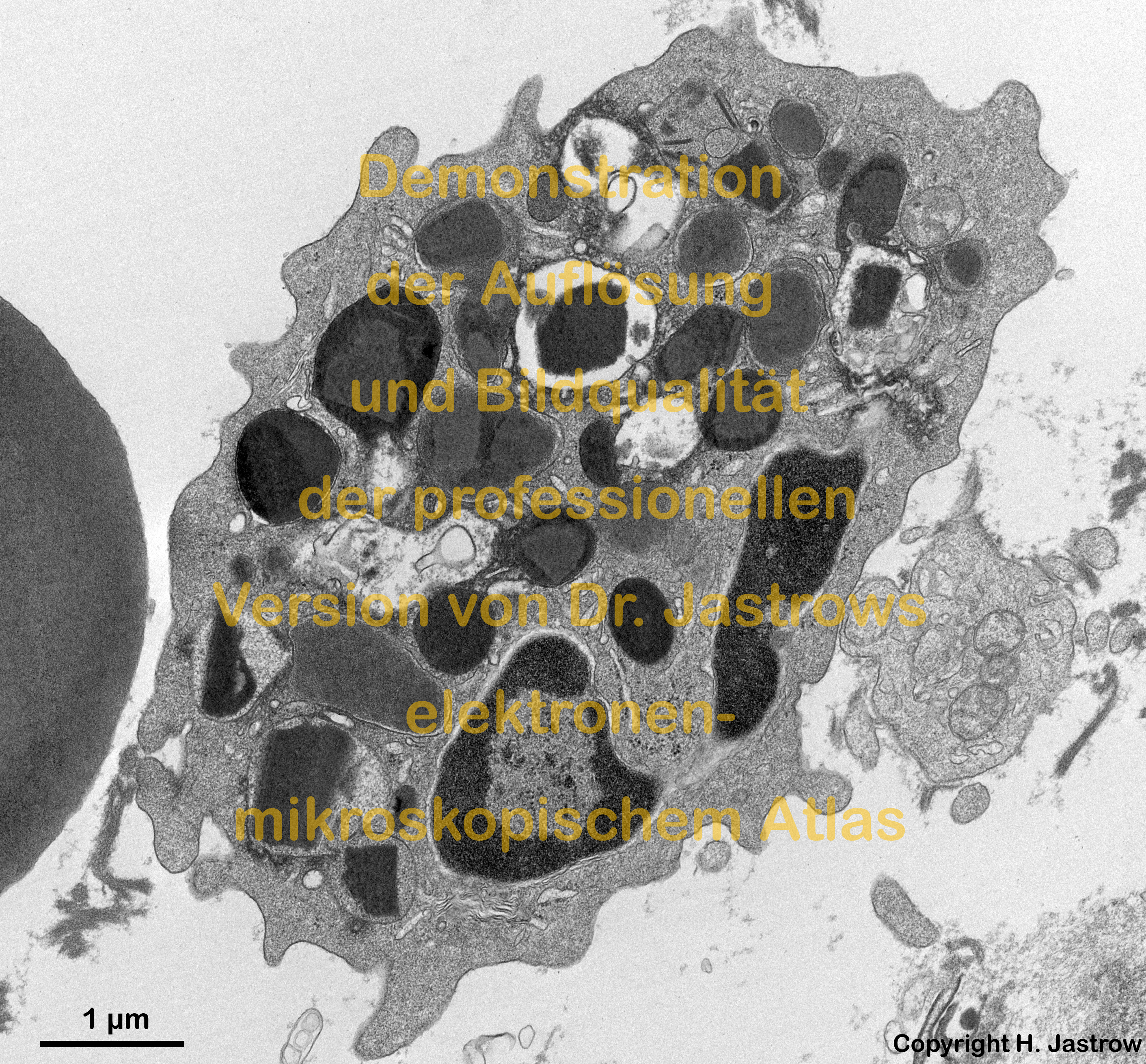
Dr. Jastrow's Light- and Electron microscopic atlas gives realistic
ideas of ultrastructures by means of over 2,200 large, high-quality
digital transmission and some scanning electron microscopic original
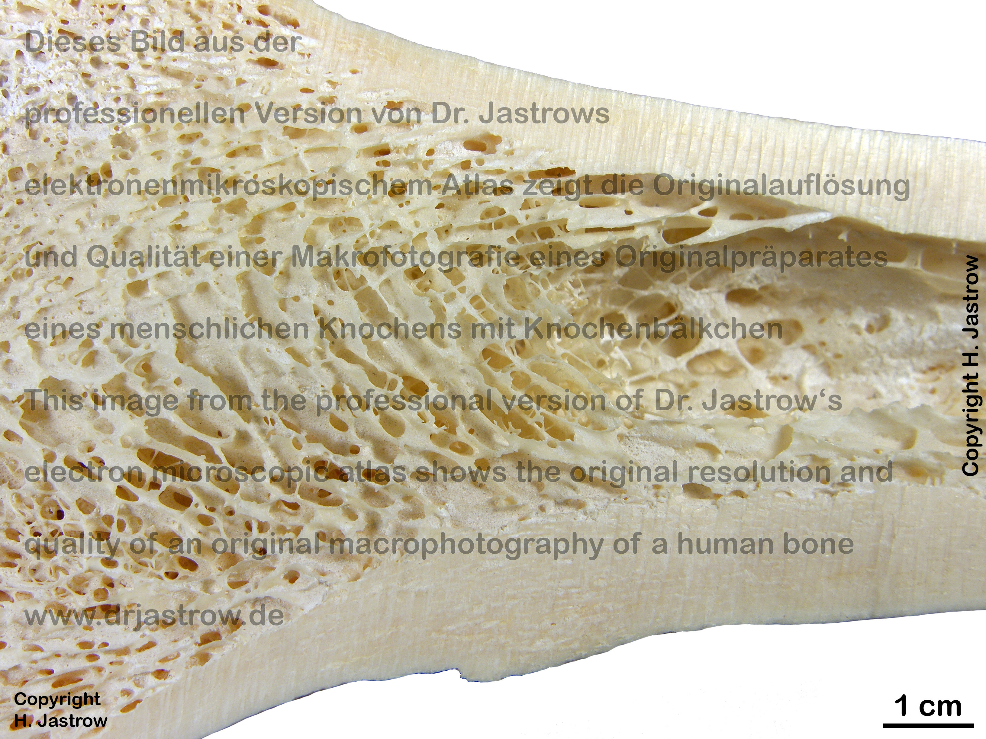
images. Further, some macrophotographies of living humans
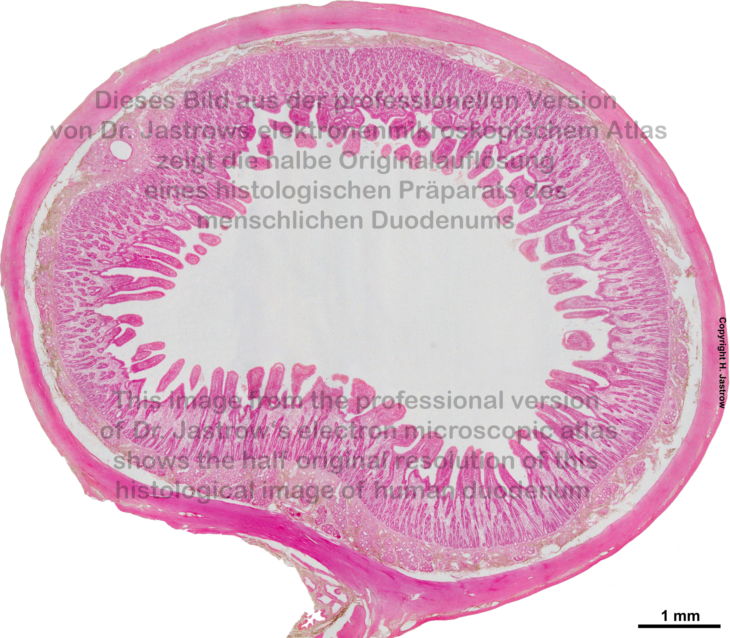
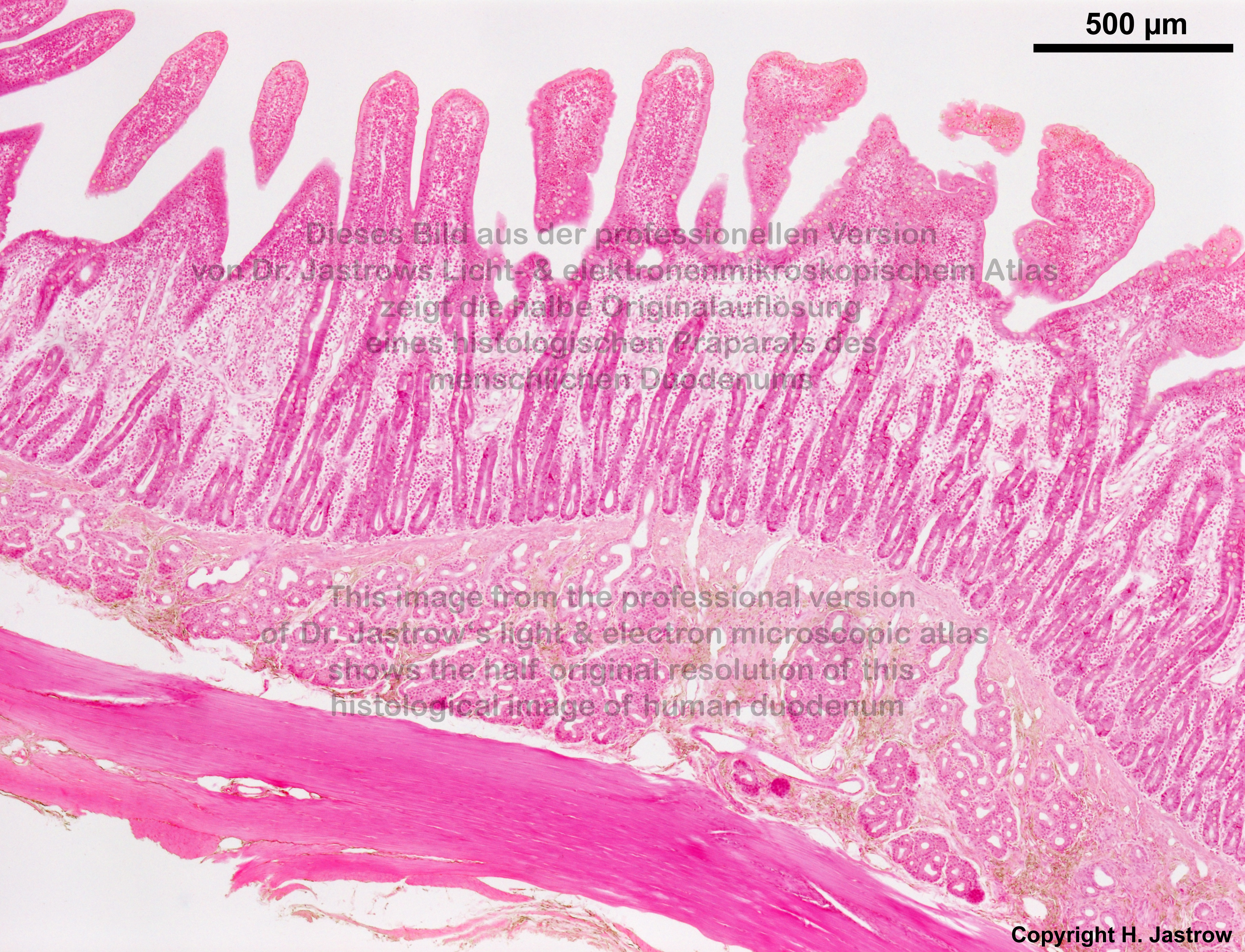
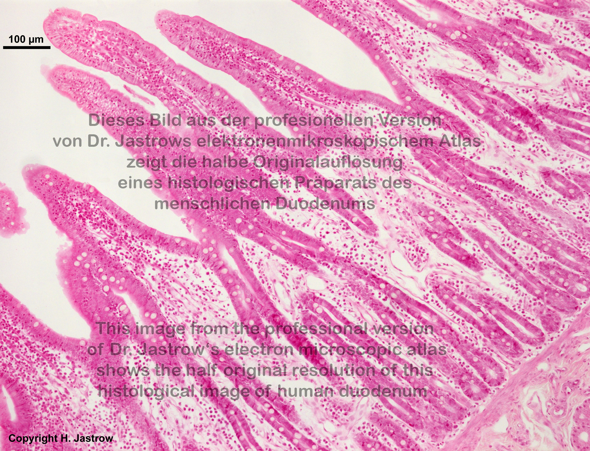
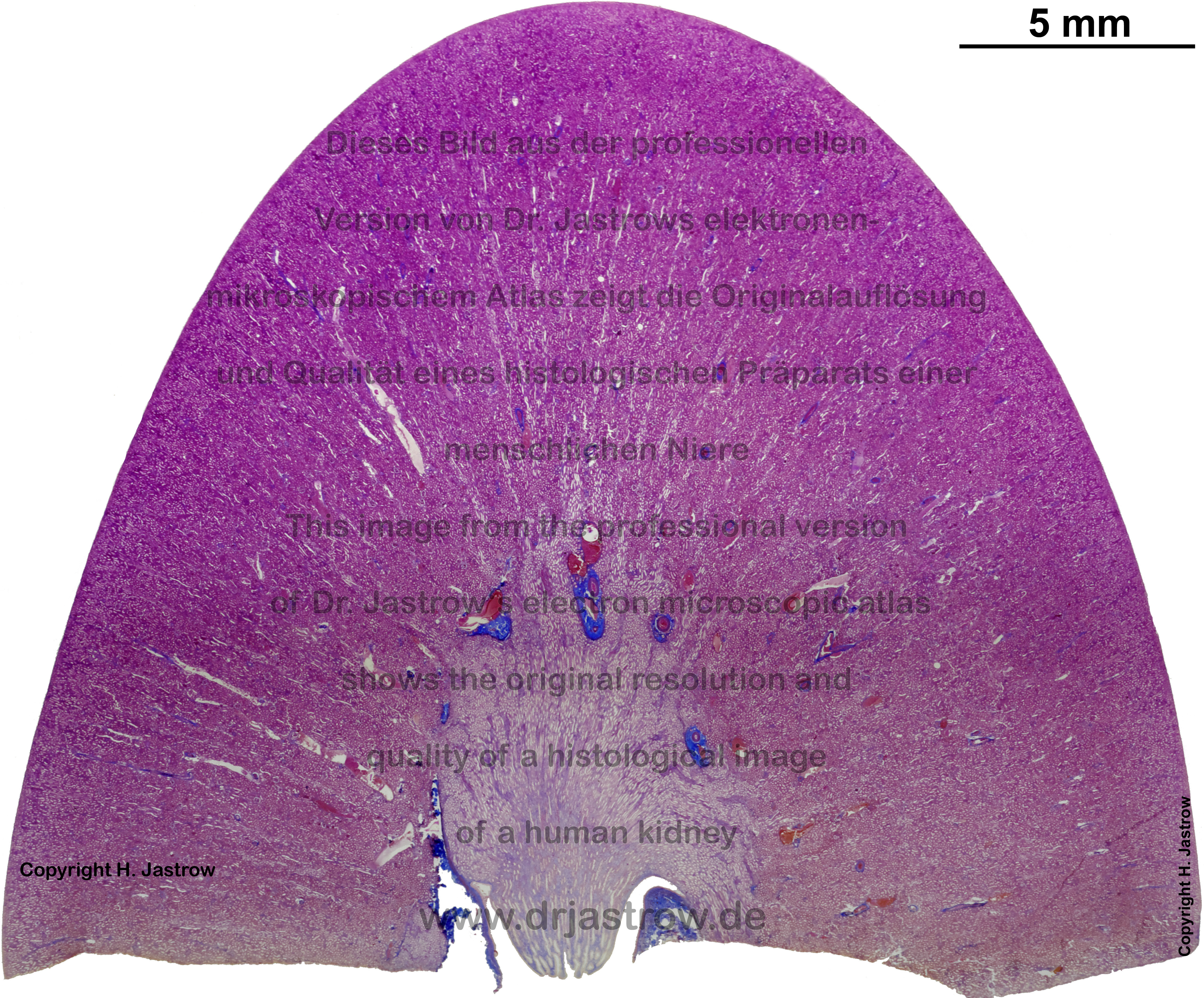
and over 2,100 light microscopic images in very high quality
and resolution allow to zoom into organs from overview via light microscopy
to fine electron microscopic detail (see
below)
The pretty complete image library of
cells,
tissues and organs
was taken from preparations of
man and/or
mammals
(monkey, rat, pig, mouse, guinea pig). Further, several images of histopathological
slides are available. The majority of pictures are multiple image aligned
digital micrographs acquired with a slow-scan CCD camera, others are high
quality scanned original micrographs or inversely scanned plate-film negatives.
Artefacts were thoroughly removed and unequal illumination was corrected
in most included images eventually additional sharpening was applied to
offer best possible quality in the digital images which are stored as maximum
quality encoded jpg files in the atlas. Choice of material is based on
quality and relevance for instruction on colleges, high-schools and universities.
Since the resolution of the images is mostly superior to 2.000
x 3.000 pixels very considerably more details are visible in comparison
to the internet version of the atlas. Every topic is covered by many different
images generally from overviews in lower magnification to very high resolution
detail pictures.
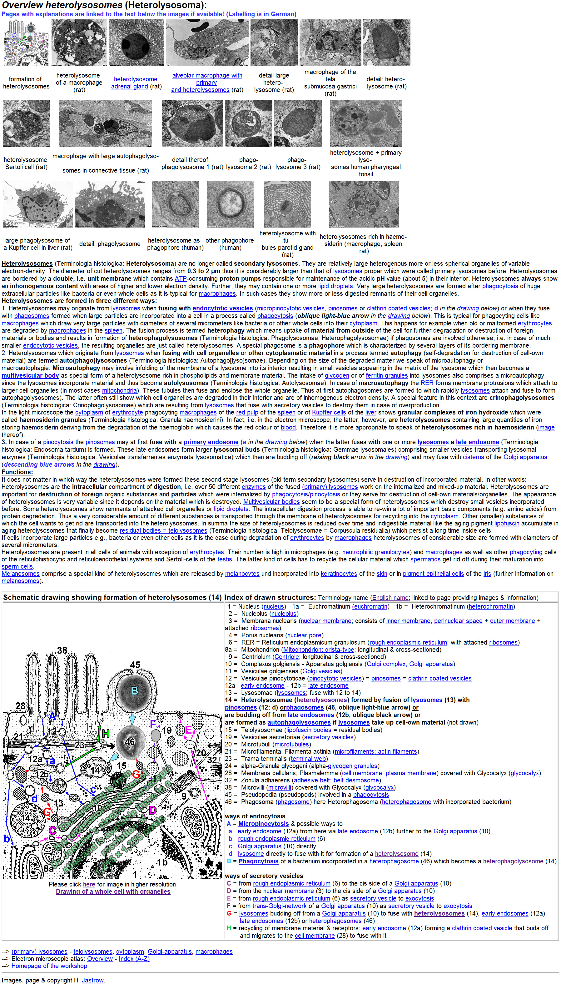
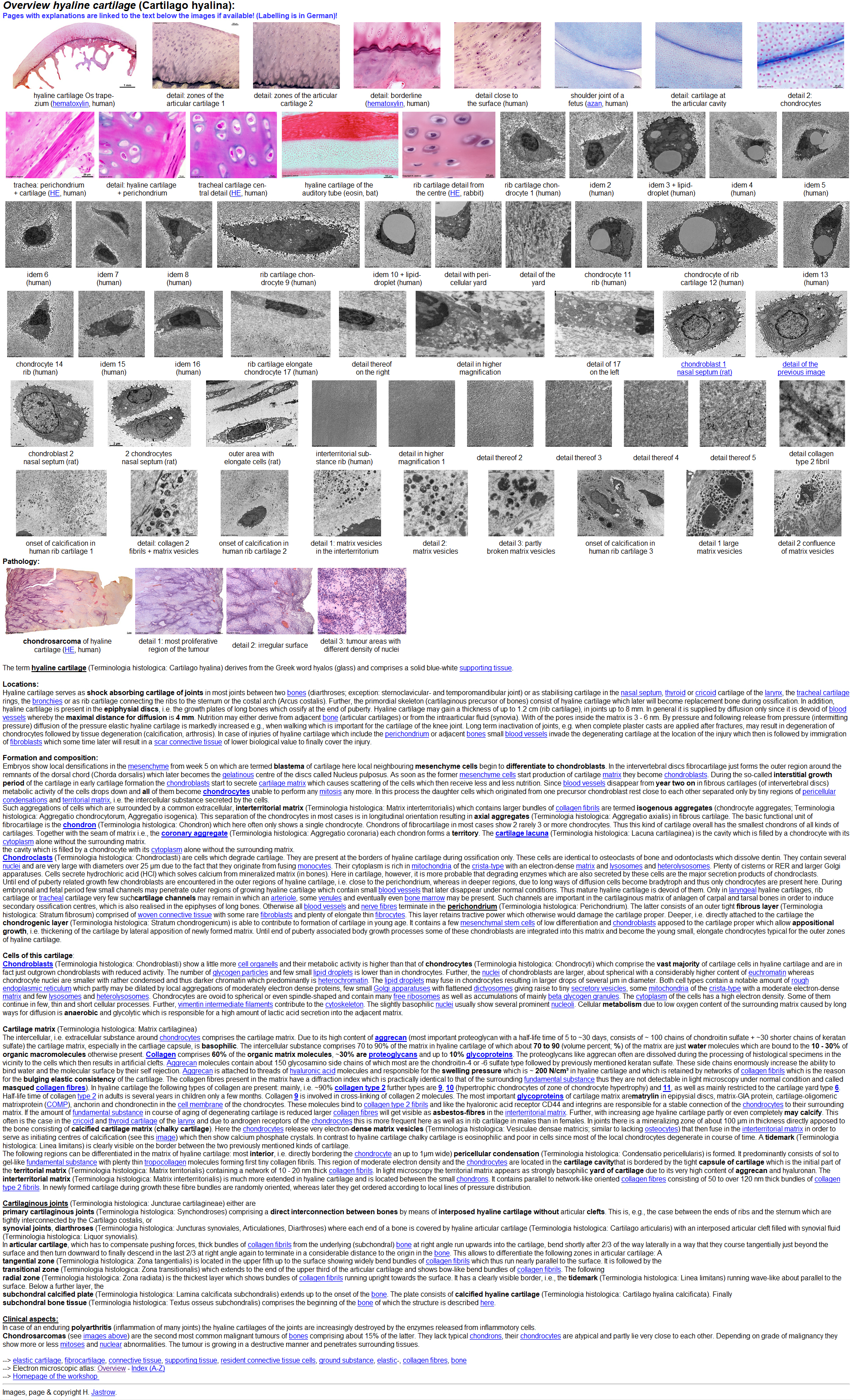
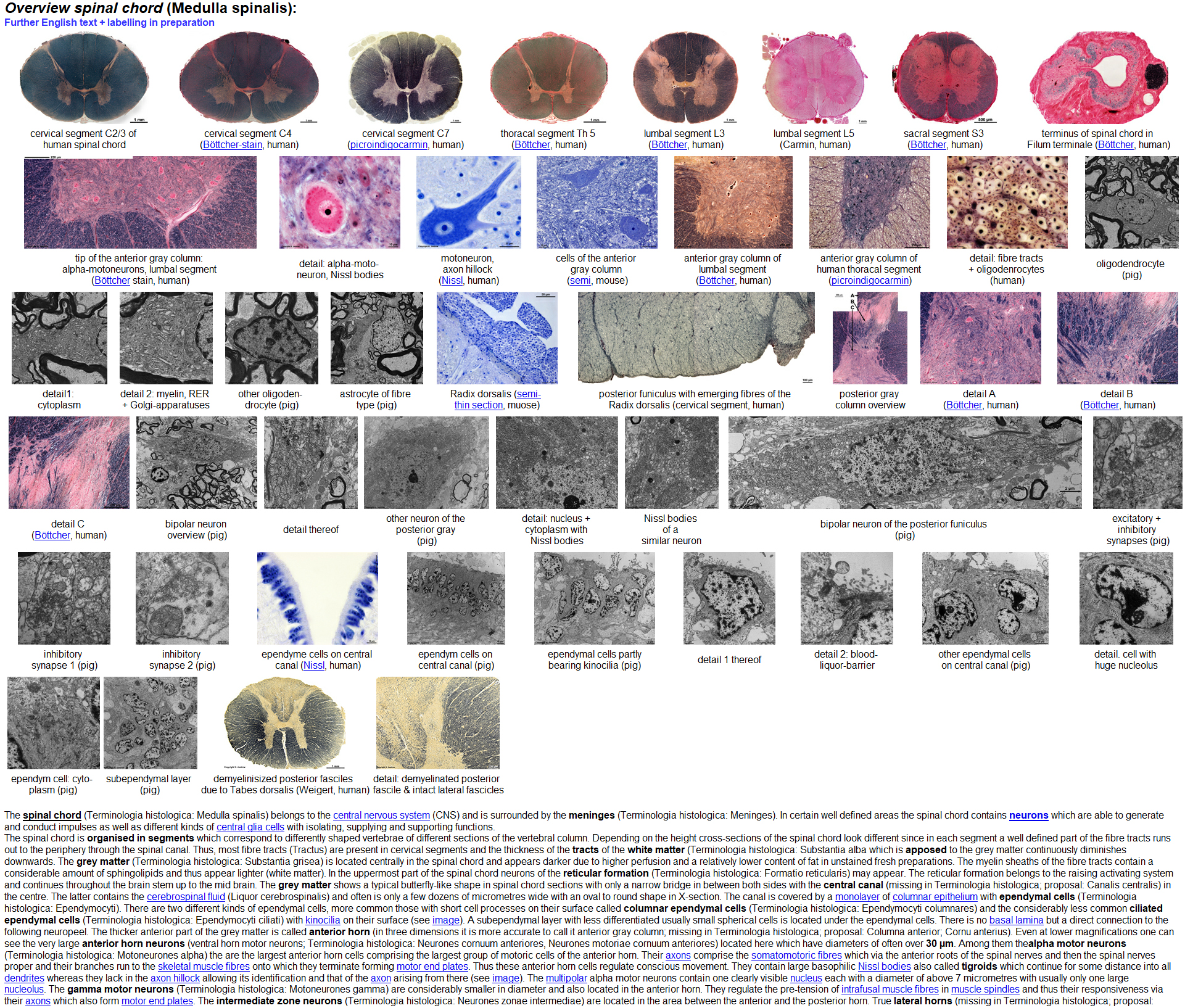
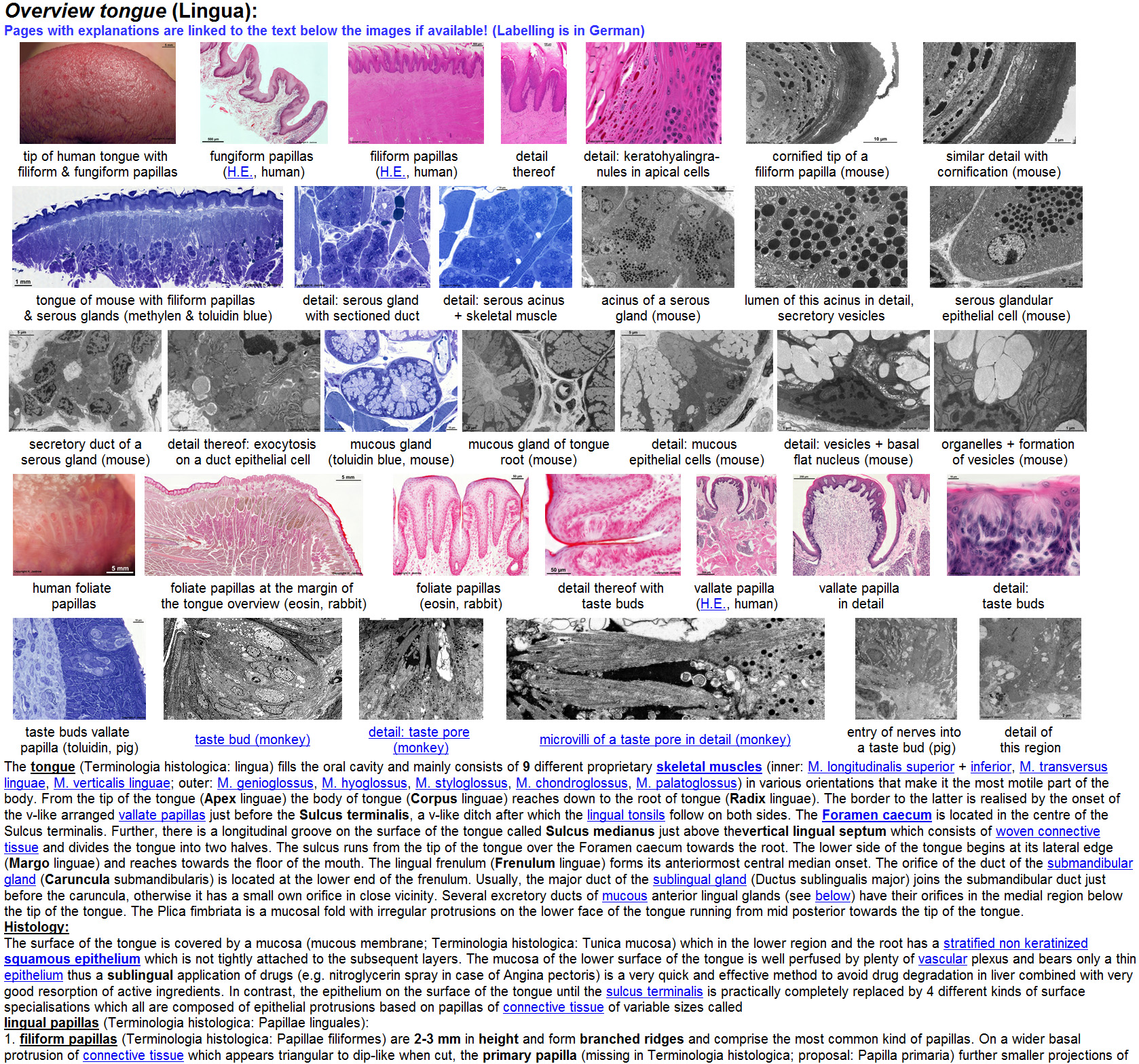
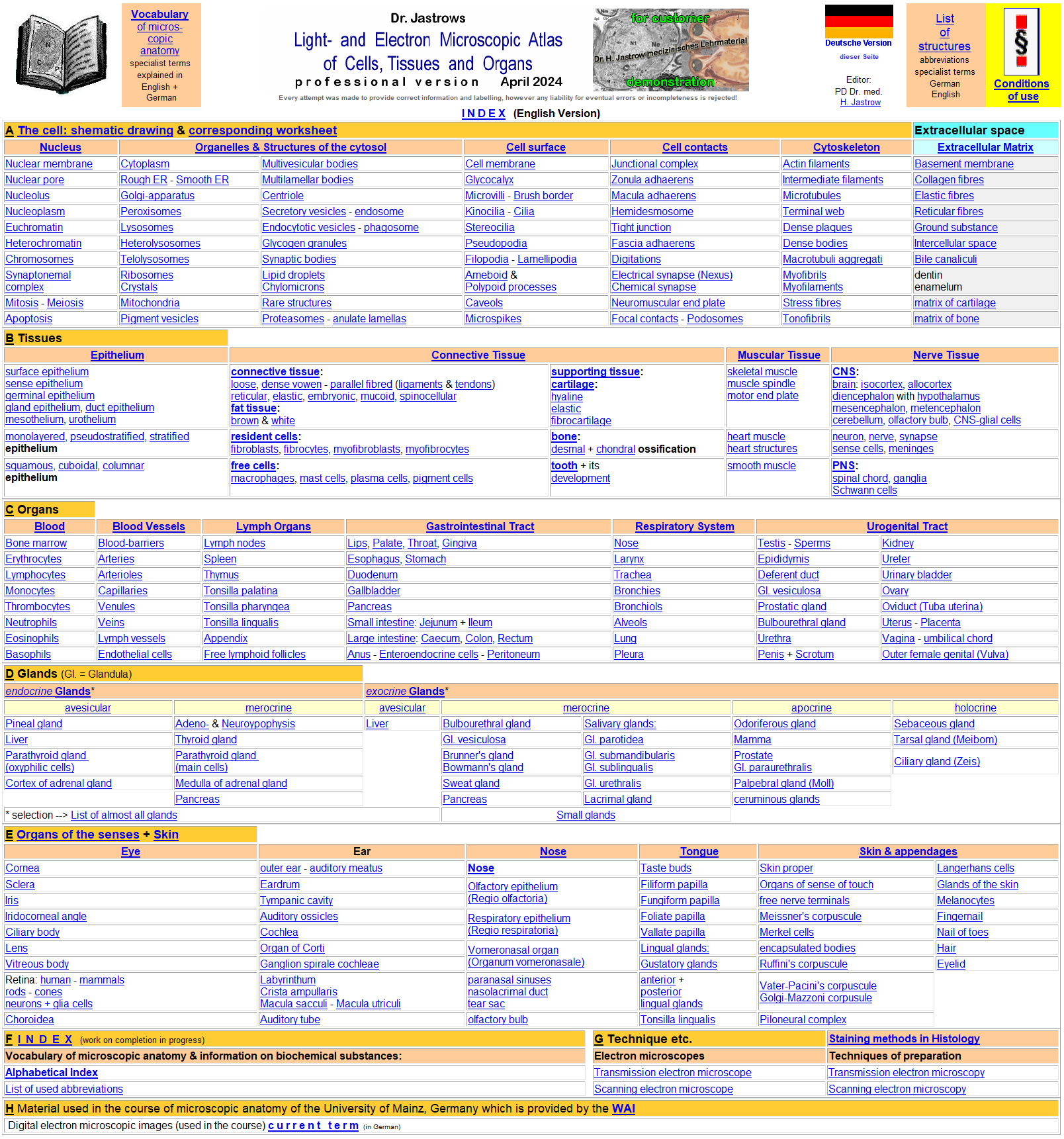
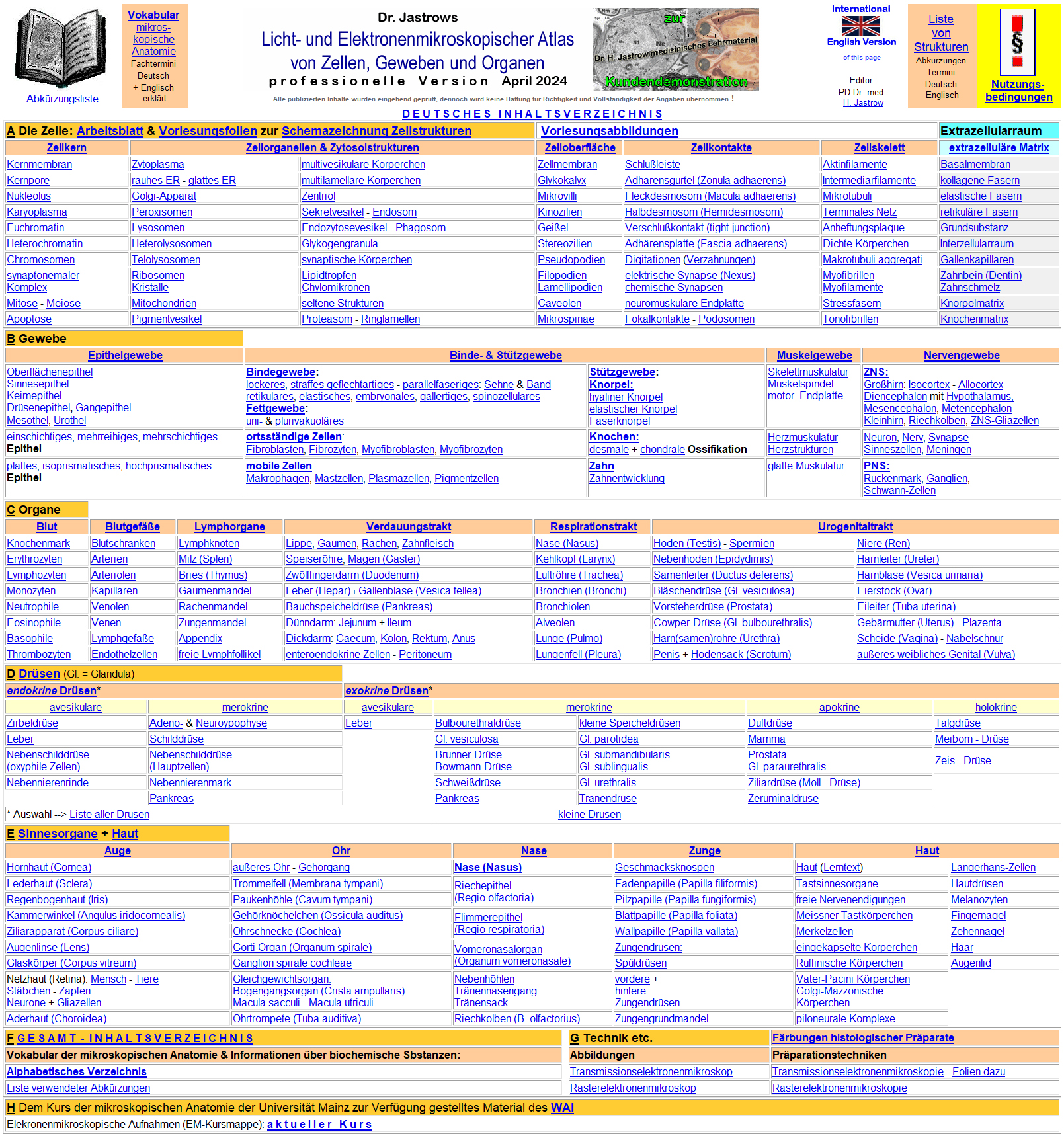
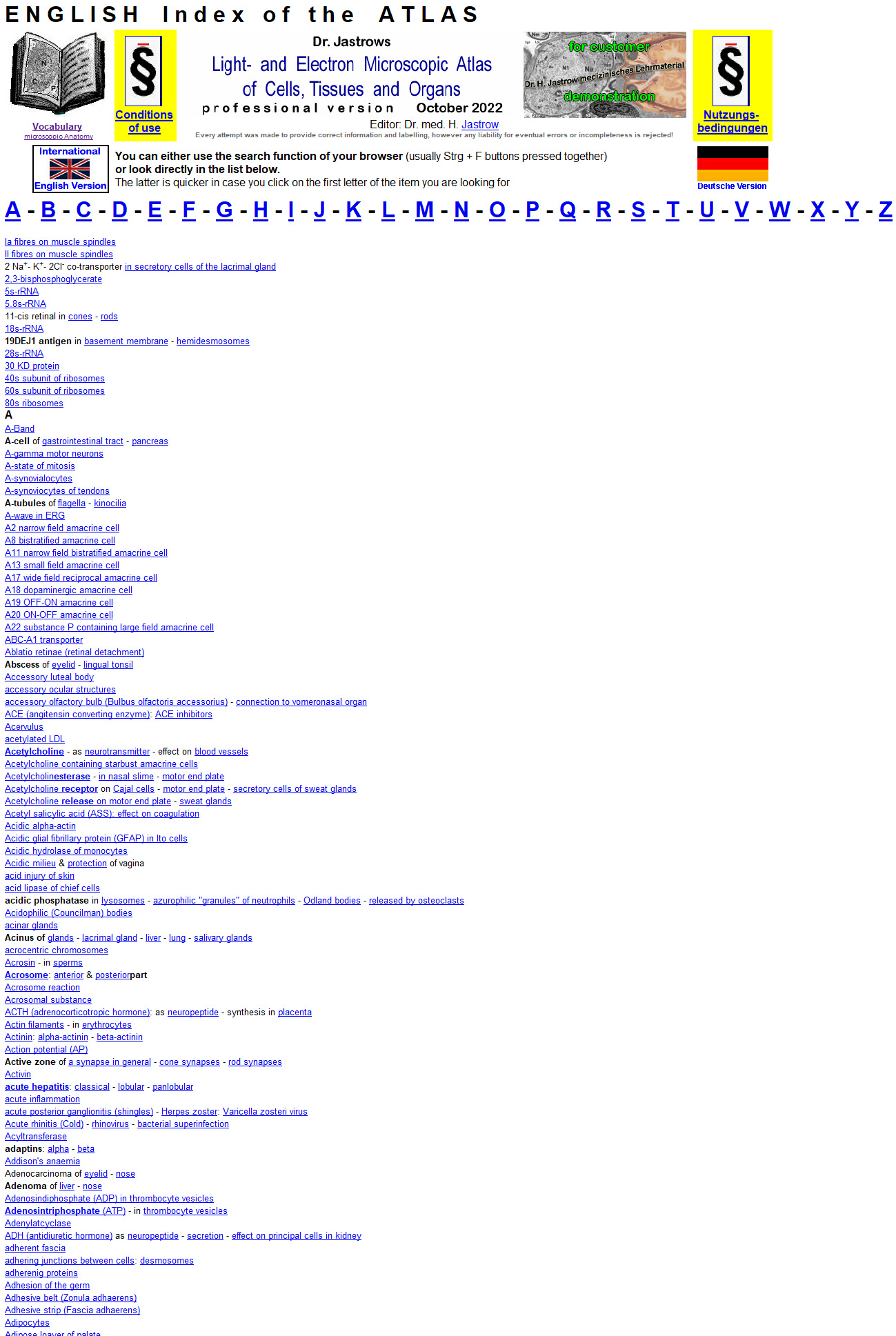
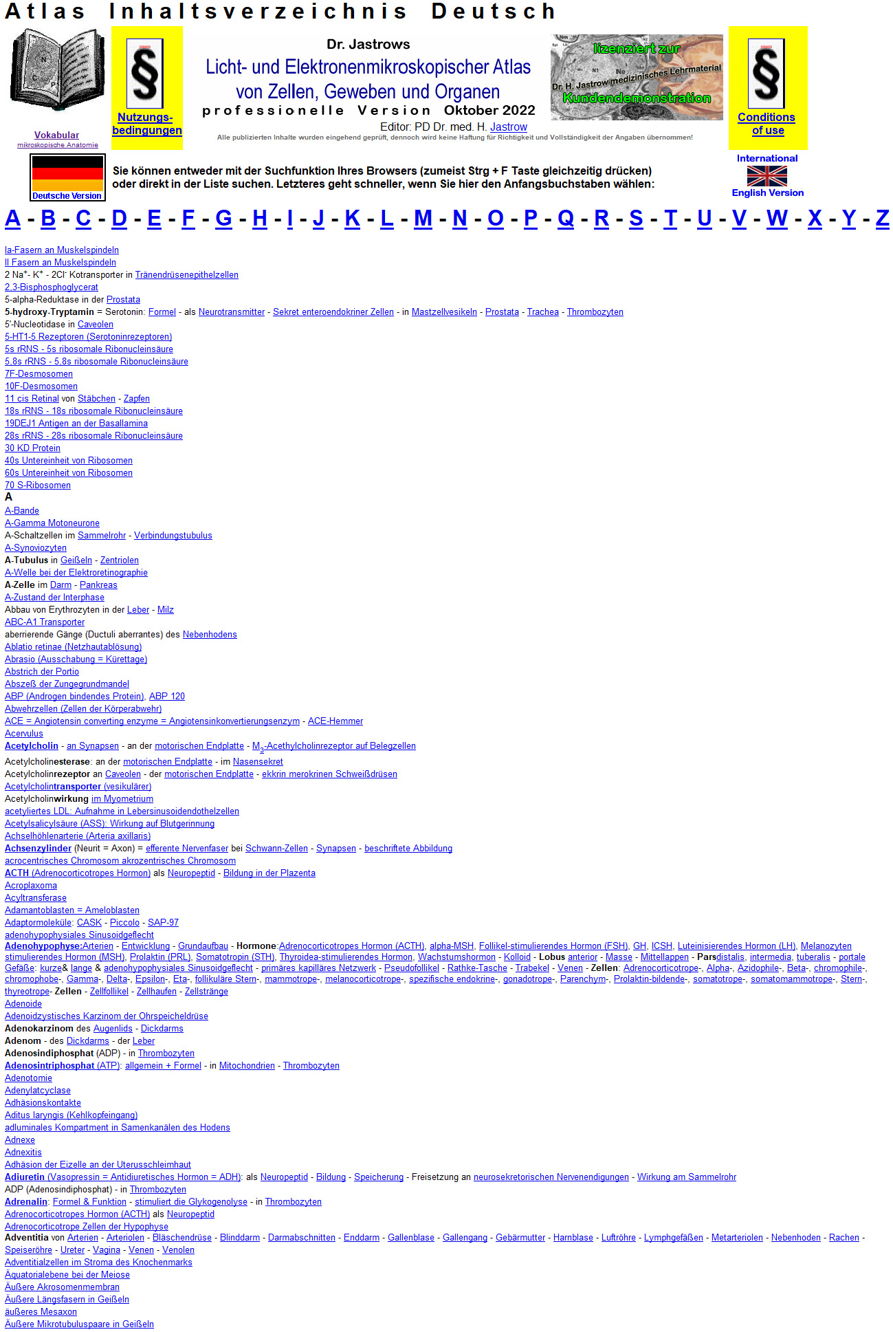
The start page (Fig.1a) in English
as well as the German one (Fig. 1b) lists important
key words which are ordered thematically. It shows an overview table
of organelles, nuclear, cellular and extracellular structures, the 4 basic
tissues, organs and organ systems. Pages providing information and image
overviews are linked to the key words. An English index page
(Fig.2a; lacking in the www-version) allows
a direct look-up of over 4,300 English terms and a German one (Fig.2b)
even >5,350 German
terms in explaining texts on the pages of the
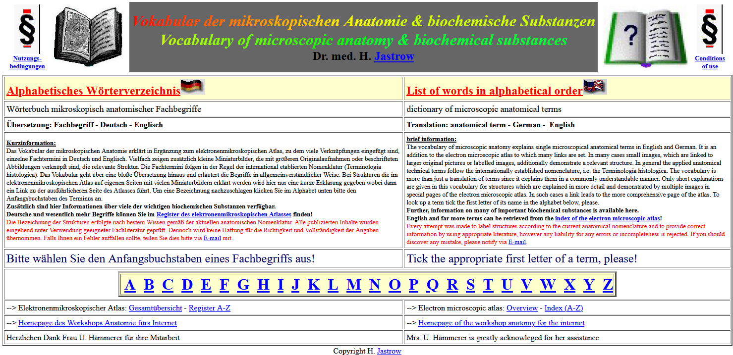
atlas. Further links form the index page lead to the main page of the integrated
vocabulary
of microscopic anatomy and biochemical substances (Fig.3,
Demo 7), providing information on vitamins, hormones
and other topics in an alphabetical choice. Further there are links to
pages explaining specimen preparation, to pages with images of transmission
and scanning electron microscopes. In addition, the overview page of the
images provided to the course of microscopic anatomy of the university
of Mainz (Germany) and a page listing the abbreviations used for labelling
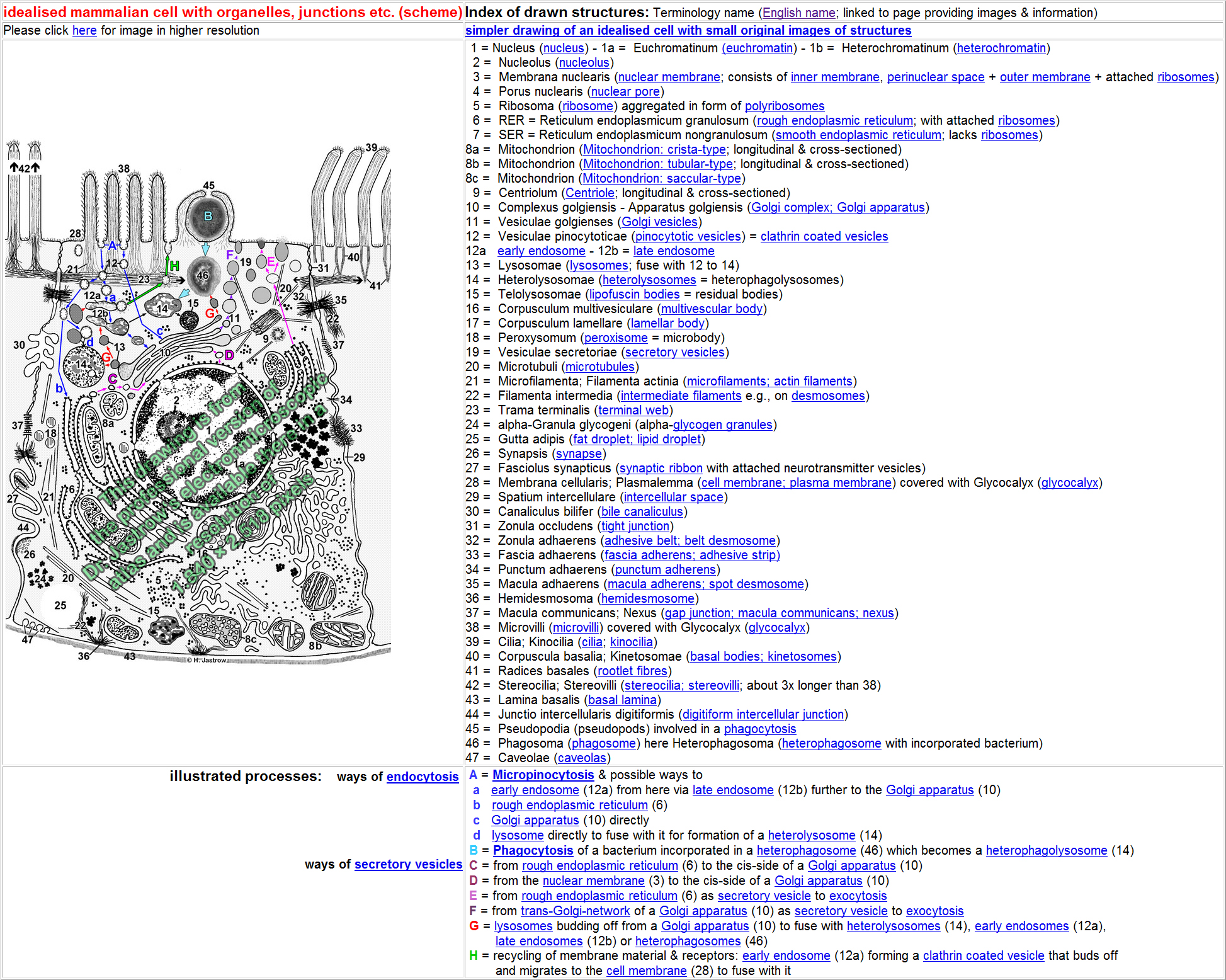
are linked to appropriate texts on the main index page of the atlas. By
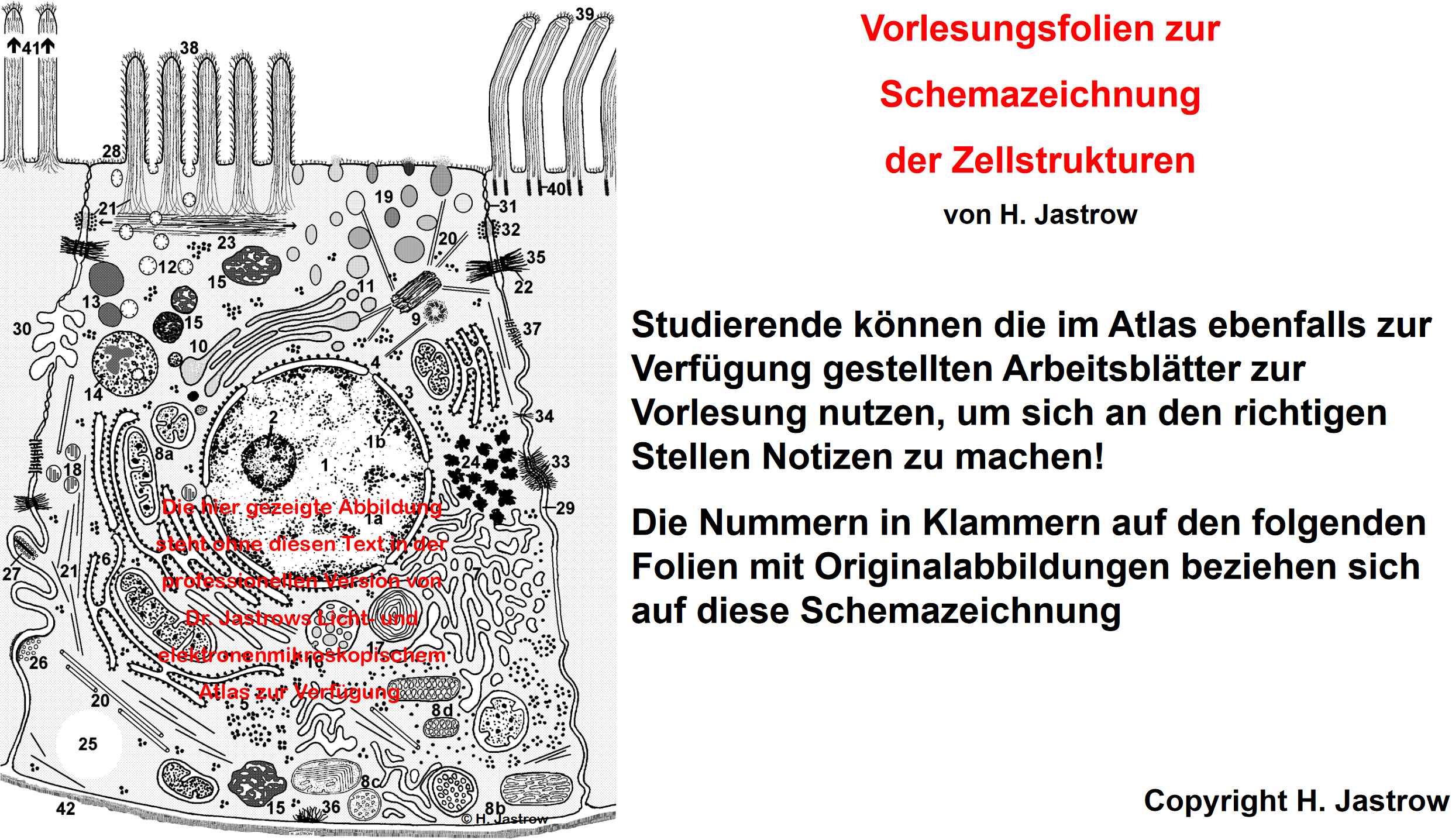
clicking on "The Cell" a page is called up which shows a schematic drawing
of an idealised cell (Fig.4) showing all
relevant ultrastructures e.g., organelles and surface specialisations.
The given names are linked to further overview pages providing appropriate
information and image overviews. The German main page further offers a
link to most instructive lecture images of cell structures with short key
words in German (sample).
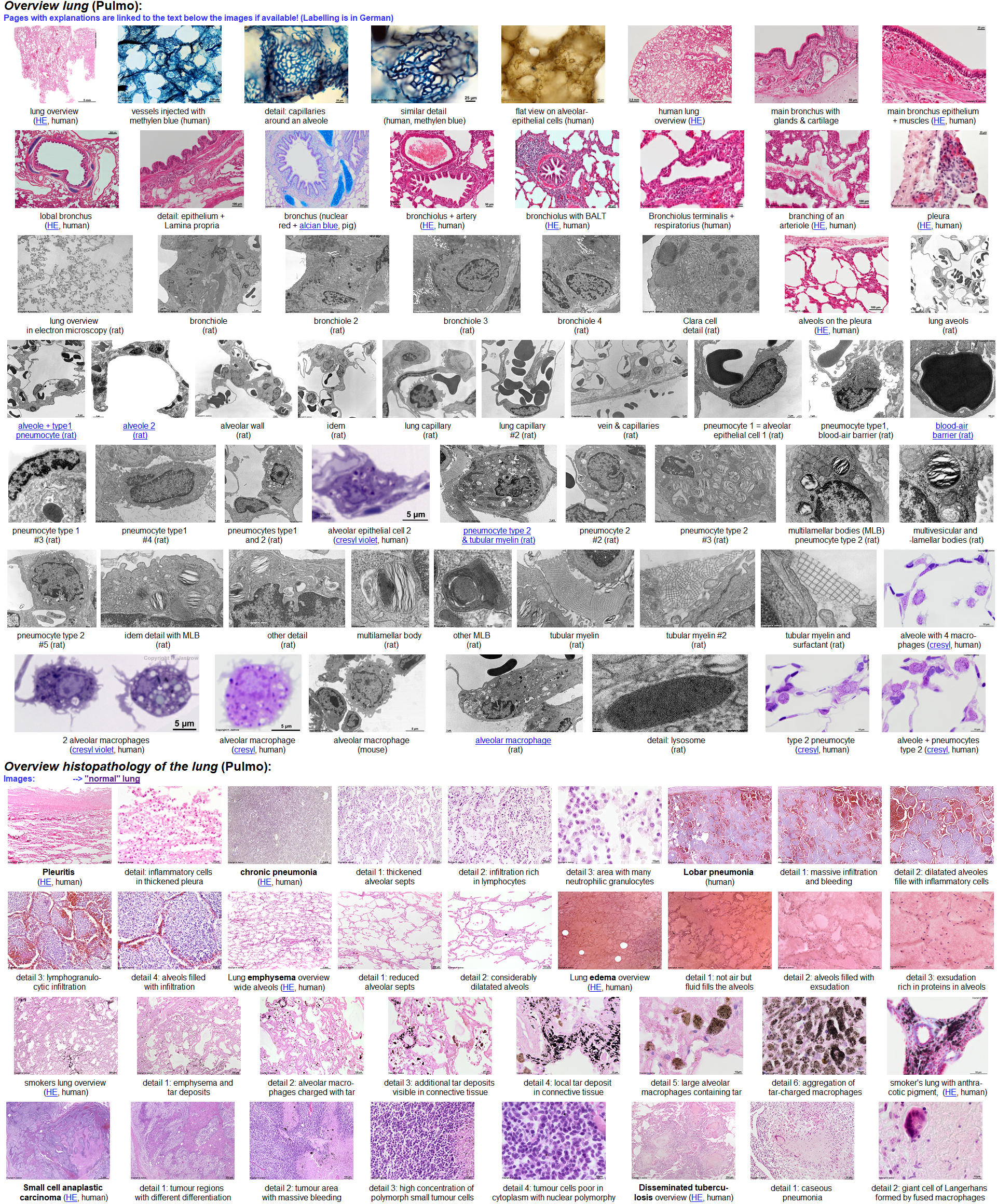
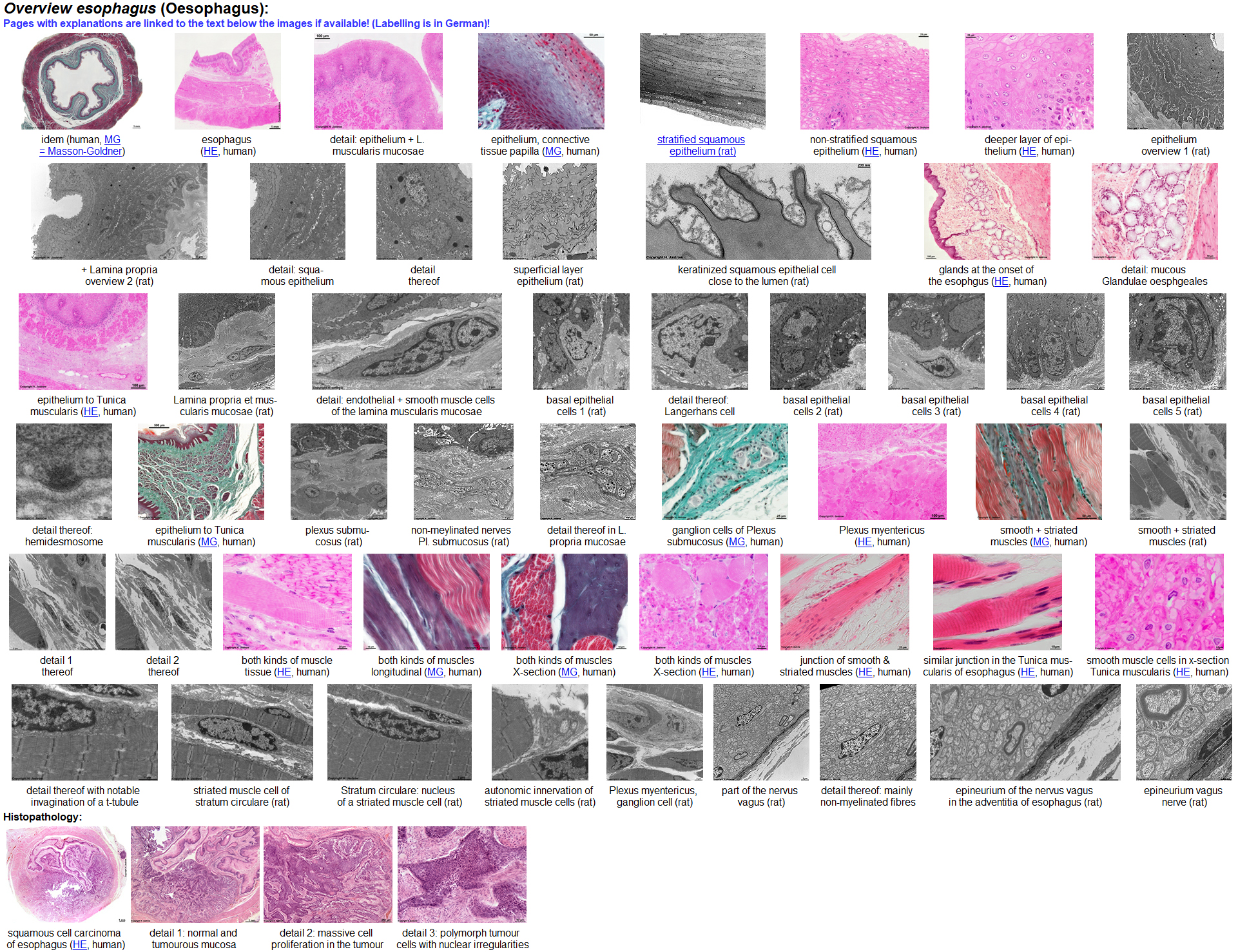
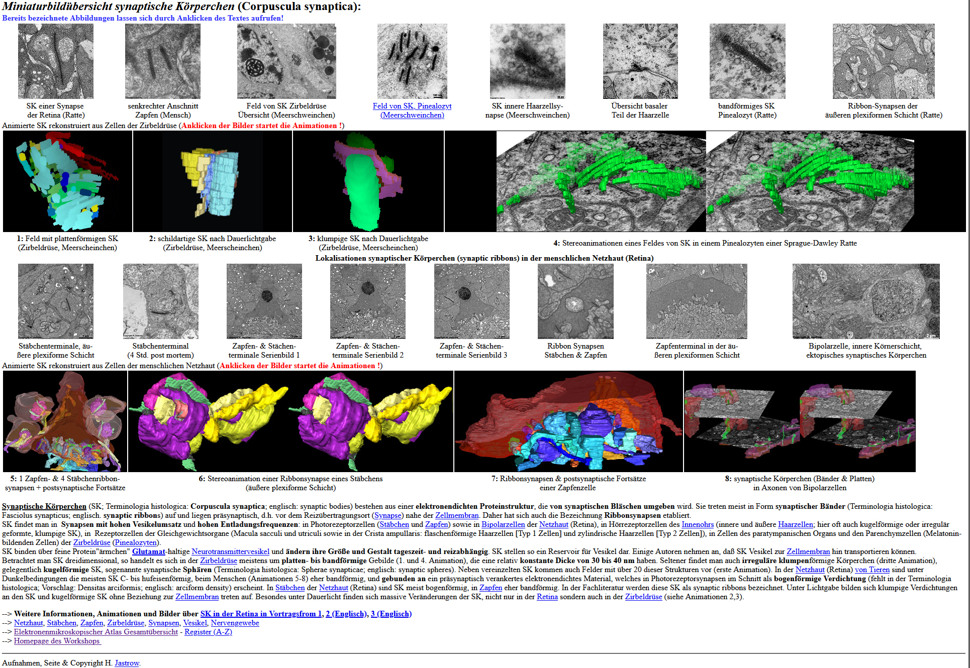
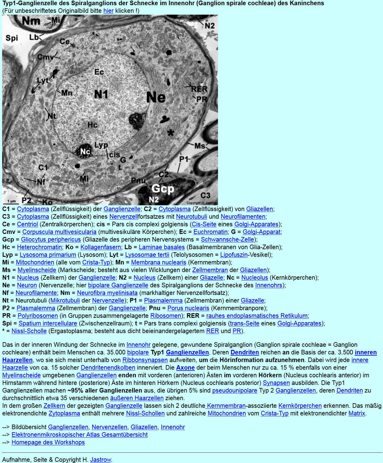
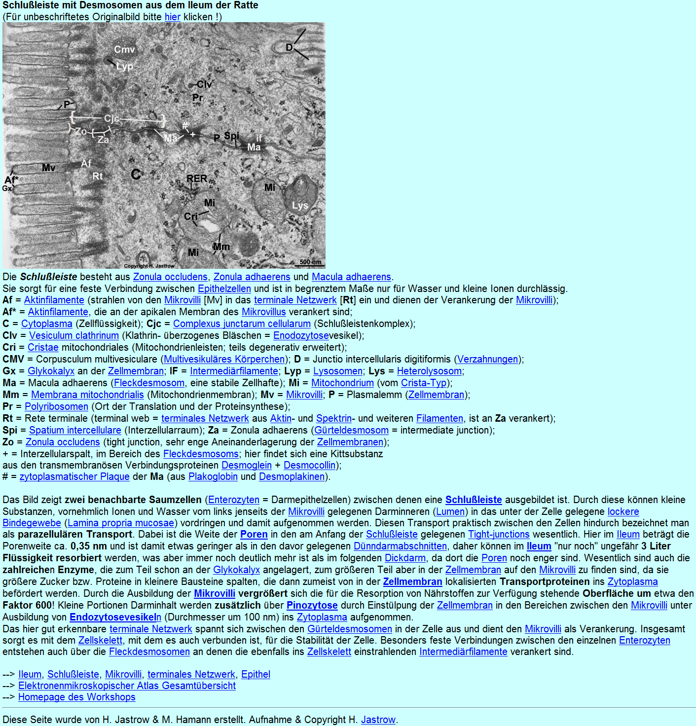
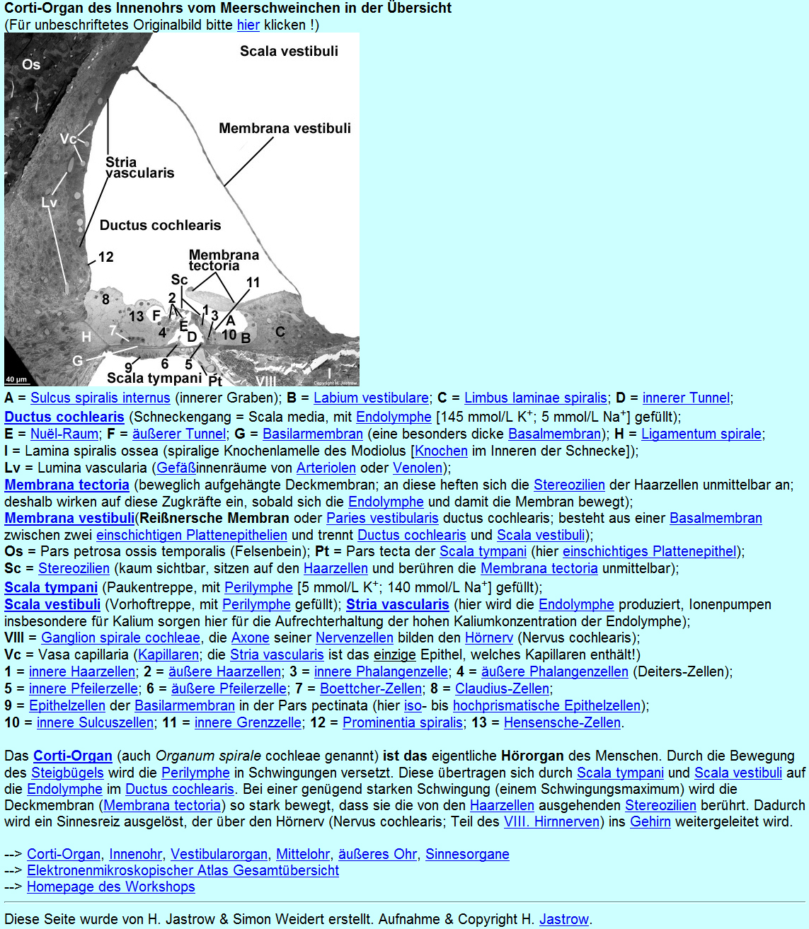
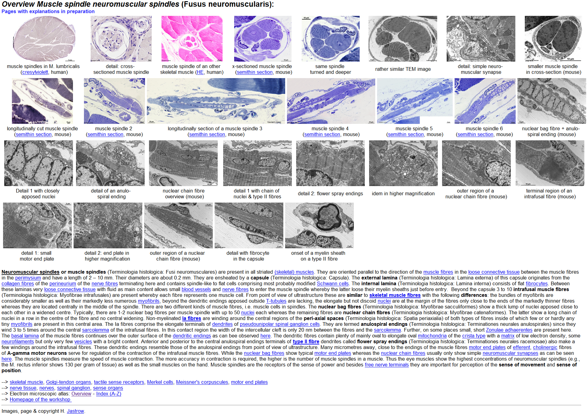
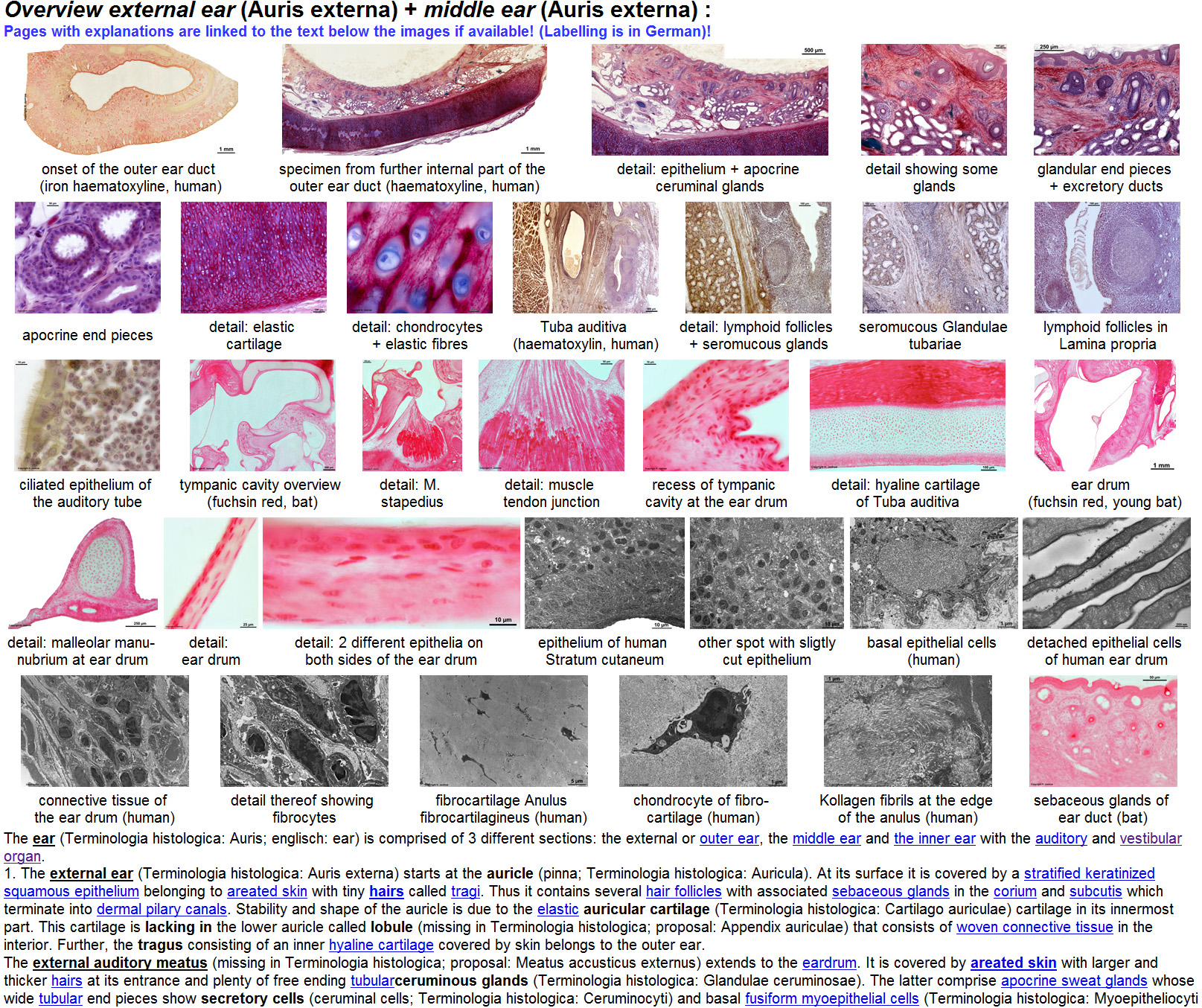
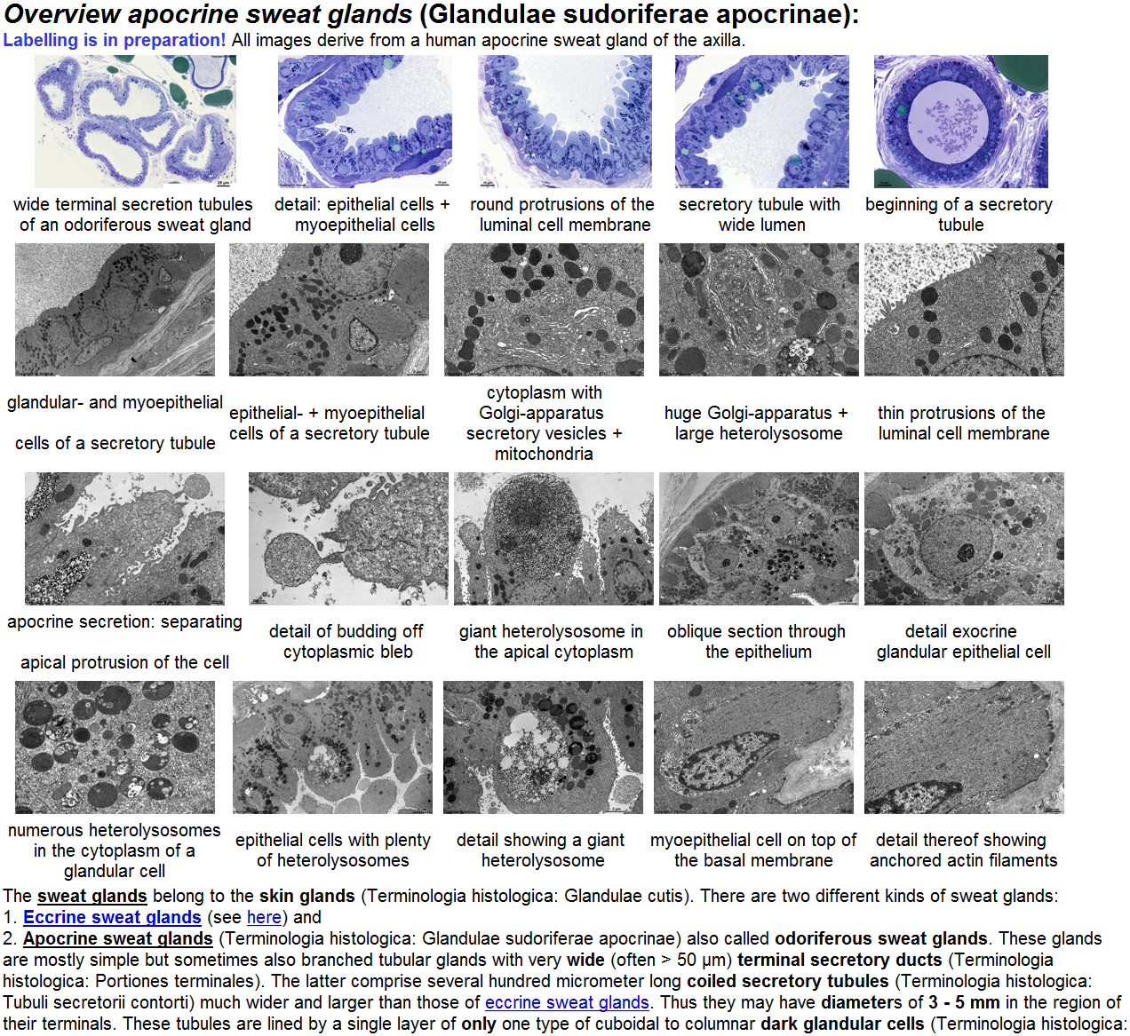
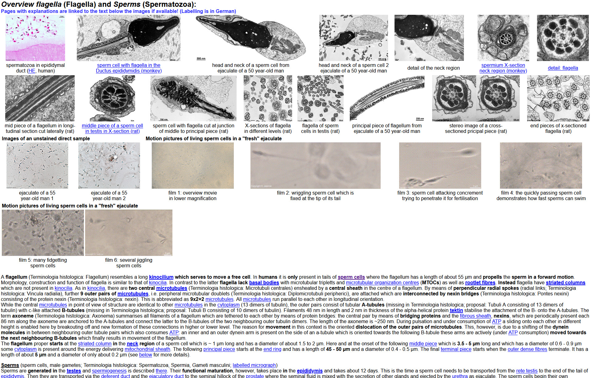
Miniature images with key word-like legends (samples) allow quick retrieval of interesting material. When clicking on an image the appropriate original image is shown as high-resolution image file (samples) providing all available details. A click on the legend of one of the over 250 labelled images leads to a page explaining the image (samples). Such pages show the whole image scaled down to a height of 500 pixels for better overview. All relevant structures are labelled with uniform abbreviations which are explained in a legend in generally intelligible way (presently in German, English translation in progress, some English pages are already available; samples). Further the atlas contains a few animations of reconstructions of retinal ribbon synapses and synaptic ribbons (sample) and 6 films of living sperms in HD resolution.
The atlas demonstrates and explains nearly all structures of cells tissues and organs. The detailed information is helpful for biology lessons in schools, colleges, high-schools and lectures, courses, seminars and other teaching events in universities. They are very use useful for basic and continued education not only in anatomy but also in all other disciplines of the biomedical research and teaching field, e.g., in pathology, molecular cell biology, biochemistry, pharmacology, physiology, clinical and biological instruction and research. The material is of value for public teaching as well as for individual learning or recapitulation. The atlas may be used as reference for "normal" ultrastructure and is a source of images and information for science, research and teaching. Selected images are used for demonstration and examination in histology courses at some places, e.g., the J. Gutenberg-University (Mainz, Germany). This choice of didactical valuable images is shown on special pages of the atlas. An intranet version installed on a password protected local network easily allows lecturers, employees, teachers and students of an institution to access the whole material as a source of images for inclusion in presentations, lectures, course or examination materials or as learning materials to understand interrelations of structure and function. Since anatomical terms and topics are explained in detail in a generally intelligible way the atlas is of value for specialist training, medical, dental and biological instruction, training in medicine associated professions, biology lessons and self-information of lay public. The labelling of structures follows the international nomenclature (Terminologia histologica) and a vast amount of (but not all) pages are available in English which allows an international use of this unique knowledge and image database. The atlas is internationally used since years by teachers of biology, high-school teachers, lecturers and medical professionals and intranet versions are used in several universities by lecturers, instructors and students. The atlas can also be used as reference of "normal" ultrastructures for comparison with own preparations. For a majority of anatomical structures a "zoom in" in many steps is possible. Where possible macrophotographies taken from living humans of a whole structure via light microscopic pictures in increasing magnification lead to images of organelles of the cells comprising the structure which are visible in electron micrographs.
The organization of the atlas allows the user to retrieve the personally relevant information interactively. Links from key words or short descriptions lead to image overviews, original images and detailed information about cell organelles in a way that a comprehensive understanding of ultrastructural interrelations is enabled. The splendid high resolution original images give a realistic picture of cell and tissue ultrastructures and in case of labelled images allow to check the own skill of knowledge. Compared to conventional often unwieldy, expensive and difficult to get comprehensive textbooks this atlas allows to see a great number of images and to get information on a topic easily using interactive link technology. Updates of this atlas which is under constant development and extension can be easily integrated and realise suggestions for improvements by users. The atlas is a detailed, extensive and easily accessible addition to other instructive material particularly since at present no comparable products are available neither in the internet nor as commercial software.
Comparison to other products:
This atlas differs from other commercial offers by its exceptional
large number of images cleaned from artefacts which are provided in very
high resolution and quality. Also the e-versions of textbooks
which I know do not provide images in comparable resolutions and very often
schematic drawings replace the true appearance of structures which can
only be demonstrated in their true nature by means of original images.
Though the atlas is not absolutely complete at present, this life-work
of over 25 years is constantly extended and optimized and
already contains "uncountable" very detailed interlinked information and
high quality images to nearly all organs and structures. Students profit
from it while learning and understanding microscopic anatomy. Thousands
of links allow to look up unclear facts quickly and it is possible to see
nearly everything that light and electron microscopy can offer. For
instructors it is easy to copy any image from the atlas and
to insert it into own presentations, e.g., lecture foils.
In this way it is possible to save the time otherwise required to obtain
images from other sources.
Perspective:
The atlas and the attached vocabulary are constantly extended
and reviewed, this includes new images and texts/text additions/corrections/updates.
It is planned to label all images in detail and to provide a virtually
complete atlas of human and mammalian ultrastructure including all kinds
of cells, organs and structures as an international reference base in
English and German. All items, topics and structures shall be explained
according to actual knowledge and shall be demonstrated by a variety of
different splendid high-resolution original images.
References:
The professional version of Dr. Jastrow's EM-Atlas is a further development
of the internet atlas of electron microscopy which was first presented
in the "Deutsches Ärzteblatt" (DÄB 98, issue 41B: 2290-2292 [12.10.2001],
as well as supplement Praxis Computer 5/2001: 17-20. Article
in PDF format). A former version was presented on the 96th meeting
of the Anatomical society at Münster, Germany on 25 March 2001 (Presentation
No. 83; Abstract published in Annals of Anatomy 183 (Suppl.), p. 50-51.
(Internet version of this presentation).
A considerable number of e-mails and server statistics of the internet
version prove a very high interest in this offer. Feedback from lecturers,
teachers, students and pupils indicate that the atlas is a very useful
and suitable aid for lecture course and exam preparation. The publication
"On the use and value of new media and how medical students assess their
effectiveness in learning anatomy" (Anatomical Record B Volume 280B, Issue
1: 20-29 [Sept. 2004]; Abstract
& link to pdf) clearly demonstrates student's demand for high quality
original image material, comprehensive explaining texts focused on the
essentials offering exam relevant knowledge and additional electronic teaching
material which is easy to handle.
Technical information and system requirements:
The atlas is provided as download and actually contains over 12.8
GB of data. After installation on a hard disk drive it may be used
with help of any common internet browser software (which is not provided
on the media) on IBM® compatible PCs. It was tested with Microsoft
Edge®, Google Chrome®, Internet Explorer®
from Version 3 and Mozilla Firefox®. Since the atlas aims
to present essentials as fast and easy as possible no complicated
java
script operations or decorative animations are applied. The atlas consists
of .html pages, jpg images and a few mp4 coded .avi animations. Thus it
is running under Windows® as well as Linux and other operation
systems. An HD resolution monitor with suitable graphic card offering a
resolution of at least 1,920 x 1,080 pixels is recommended since
the atlas is optimized to this resolution. Otherwise only very small
parts of the large original images are visible when looking in original
resolution.
Demo version on the internet:
The atlas is published freely accessible in the WWW in a considerably
reduced manner (image resolution and quality are very significantly
lower, NO index page, NO macrophotographies, NO images from histology).
Please note that the www version has NOT been updated since December
2013. For this reason the presently over 100 new pages (see
below)
and all updates of the professional version, which is under constant further
development, are NOT available online.
To examine the www-version use this link, please: http://www.drjastrow.de/WAI/EM/EMAtlas.html
I am happy to answer your further
questions:
Holger Jastrow
| PD Dr. med. Holger Jastrow
homepage address E-mail: info@drjastrow.de |