Overview Golgi-apparatus (GA;
Complexus golgiensis; Apparatus golgiensis):
Pages with explanations are linked to the
text below the images if available
 |
 |
 |
 |
 |
 |
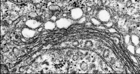
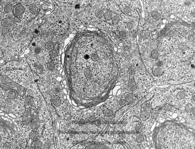
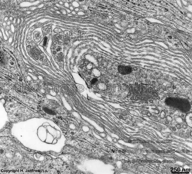
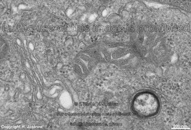
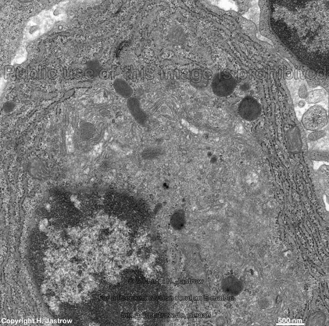
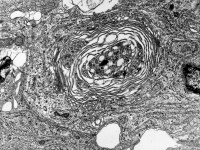
round GA of an epthelial cell of
the epididymal duct (rat) |
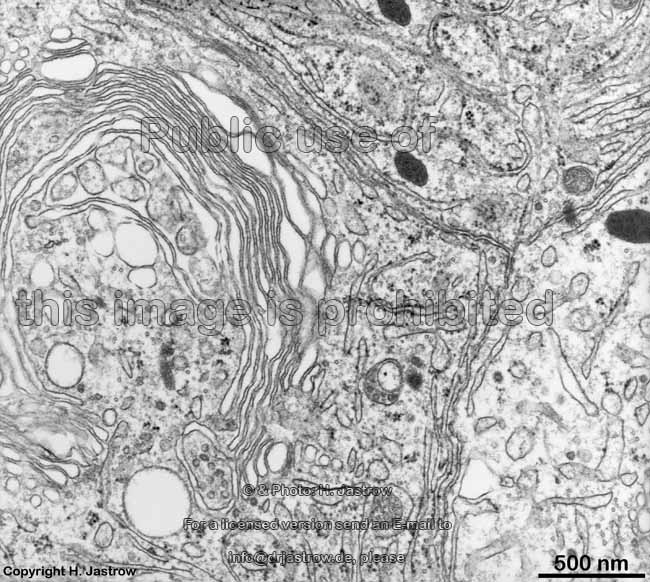
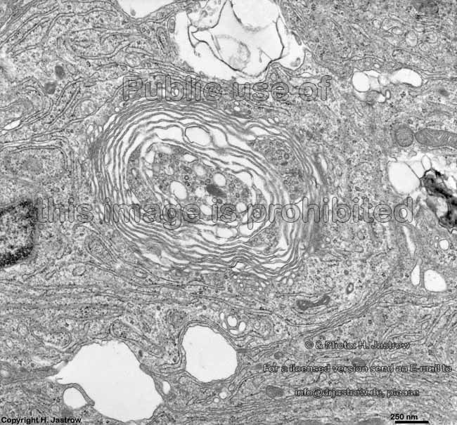
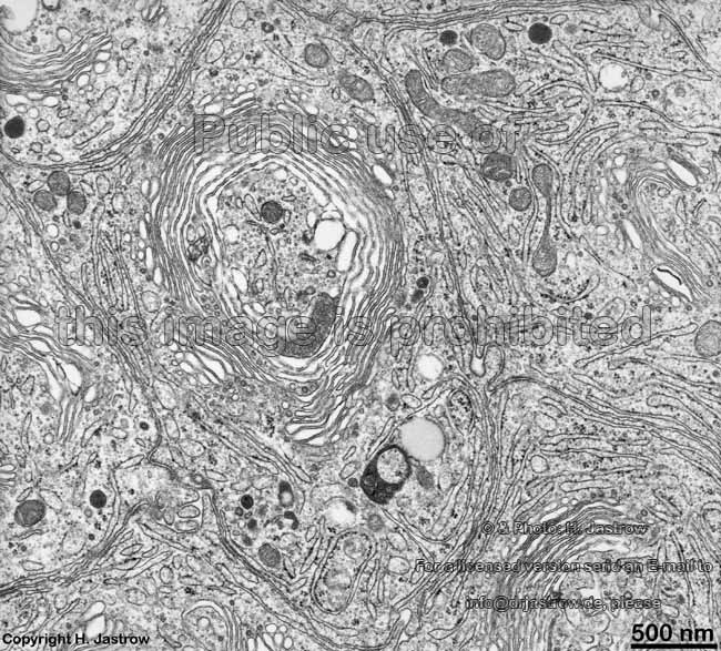
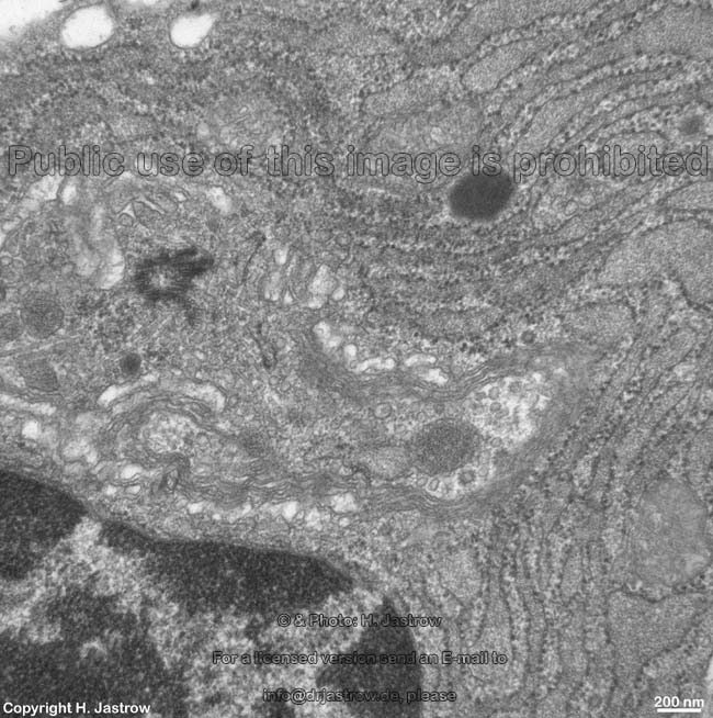
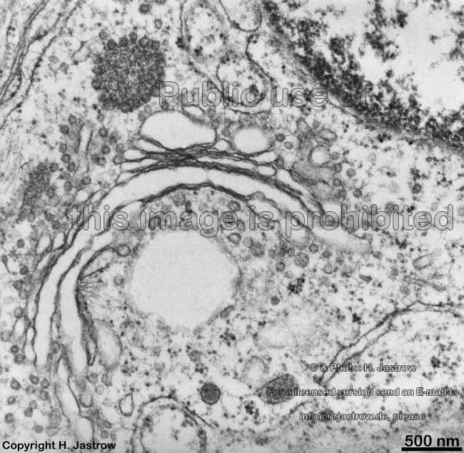
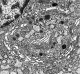
GA of a Bowmann
gland of the nose (monkey) |
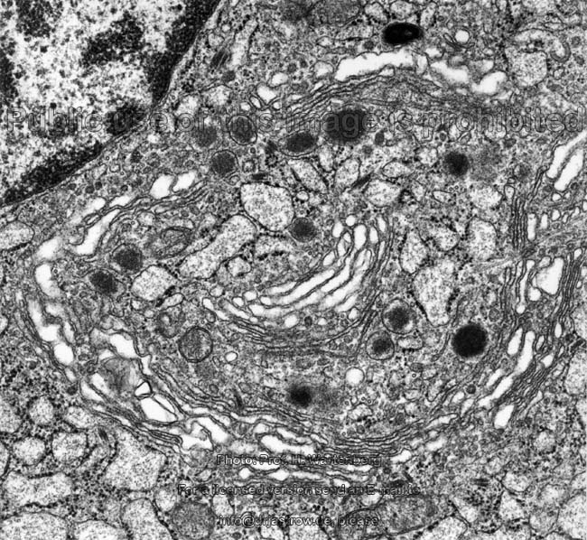
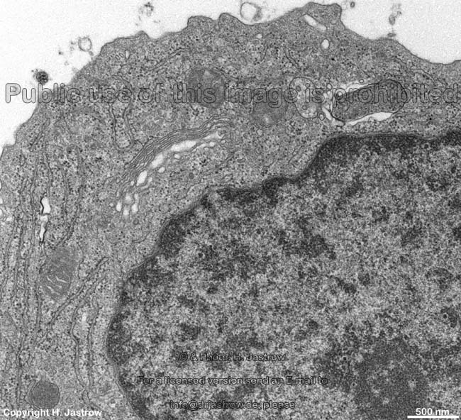
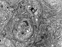
GA od an epithelial cell of
the epididymal duct (rat) |
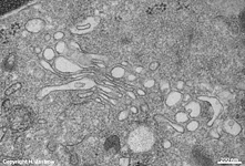
GA of an epithelial cell
of the epididymal duct (rat) |
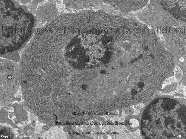
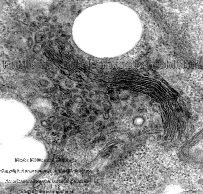
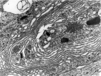
Stereo-image od a GA of a supporting
cell of the organ of Corti (guinea-pig) |
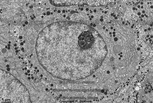
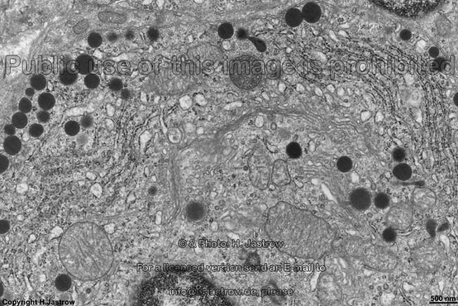
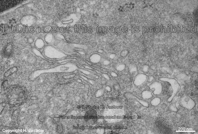
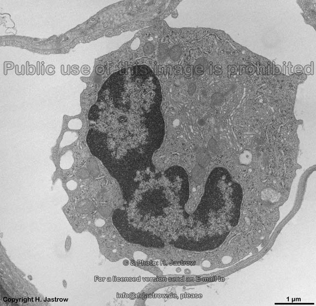
Golgi-apparatus, neutrophi-
le granulocyt (human) |
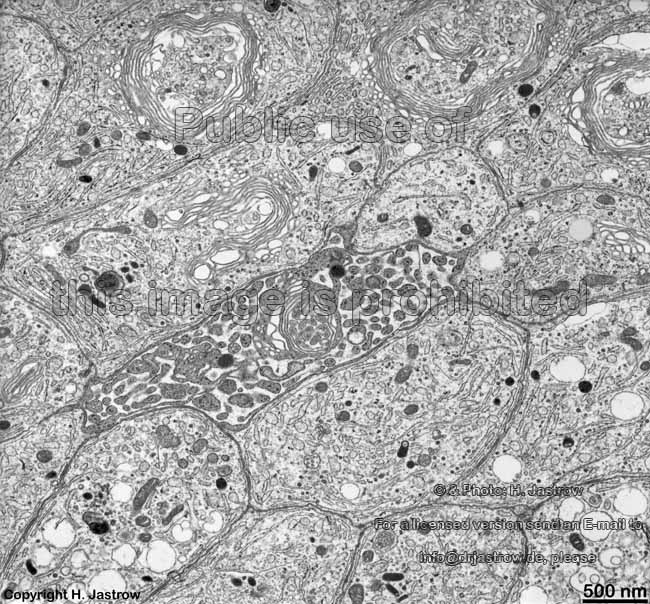
The Golgi apparatus (Terminologia histologica: Complexus
golgiensis or Apparatus golgiensis) is a lamellar membranous structure
near the
nucleus of almost all cells. It consists
of curved parallel series of flattened saccules called dictyosomes
that are often expanded at their ends. In secretory cells e.g., salivary
glands, the apparatus concentrates and packs the secretory products.
The GA serves for regeneration of the cell
membrane (plasmalemm) and modification of proteins (e.g. joining of
proteins and glucuronic acids). Vesicles of
the RER fuse on the cis-part of the GA
while so called Golgi-vesicles are released on its trans-part. The
so called cis side of the GA is oriented towards the rough
endoplasmic reticulum (RER) whereas the opposite trans side
gives raise to the secretory
Golgi vesicles. The distance between the dish-like cisterns of the GA.
which at their edges show sac-like protrusions giving raise to small vesicles,
is only 20 - 30 nm. These small vesicles shall mainly serve for transport
of material from one to the subsequent disc that always is from cis
to trans. If these vesicles contain enzymes in high concentration they
are termed
primary lysosomes. In case they contain
smaller vesicles, they are called multivesicular
bodies.
The name of the organelle derives from Camillo Golgi (1843 - 1926),
a pathologist who worked in Parvia. In the light microscope GA are hardly
visible as light areas close to the nucleus
at highest magnifications. GA may be stained using osmium tetroxide or
silver salts for light microscopic investigations. The perinuclear
(= near the
nucleus) location is typical
for round cells. In the secretory
epithelial cells of glands the GA is
always in a supranuclear position, i.e. above the nucleus
on top of more or less RER in direction to the
apical cytoplasm following the way of
production of secretion which finally is released from the uppermost Golgi-vesicles
into a lumen. In many cases two larger GA arrange around the 2 centriols
which is due to microtubules originating
there. These microtubules shall serve for
stabilisation of the GA and for its positioning in cells. In general the
GA is present in all cells bearing a nucleus
and its size depends on cell activity.
--> cytoplasm, RER,
SER,
vesicles,
endocytosis,
primary
lysosomes, peroxisomes,
multivesicular
bodies
--> Electron microscopic atlas Overview
--> Homepage of the workshop
Two pictures were kindly provided by Prof. H. Wartenberg,
one by Dr. M. Klinger; other images, page & copyright H. Jastrow.