Overview secretory vesicles (Vesiculae
secretoriae):
Pages with explanations are linked to the
text below the images if available! (Labelling is in German)
 |
|
|
|
|
|
|
|
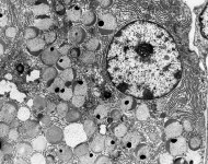
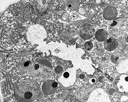
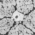
submandibular gland
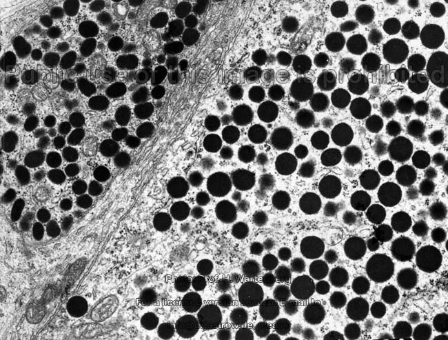
serous acinar cell (monkey) |
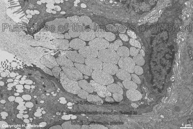
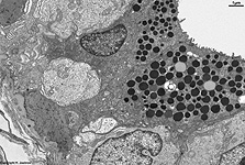
acinus with serous se-
cretion vesicles 1 (monkey) |
acinus with serous secre-
tion vesicles 2 (monkey) |
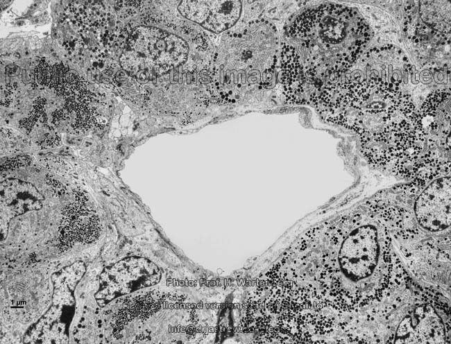
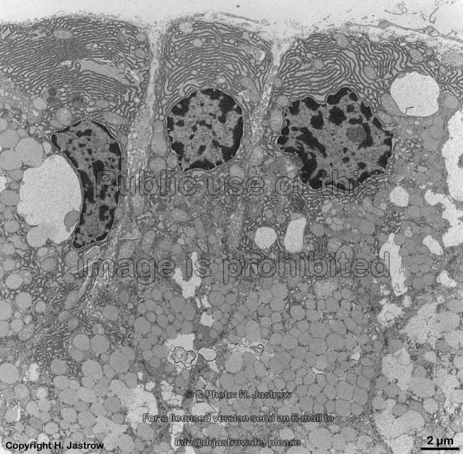
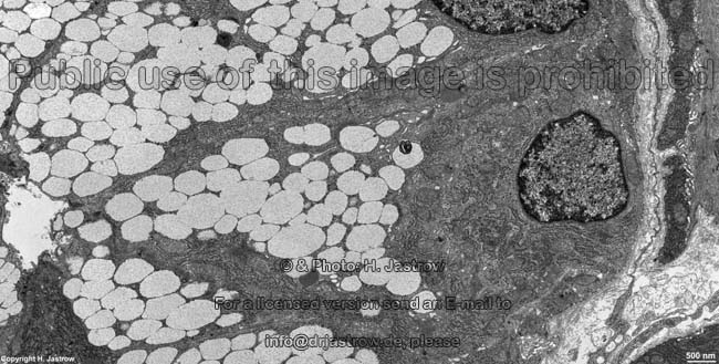
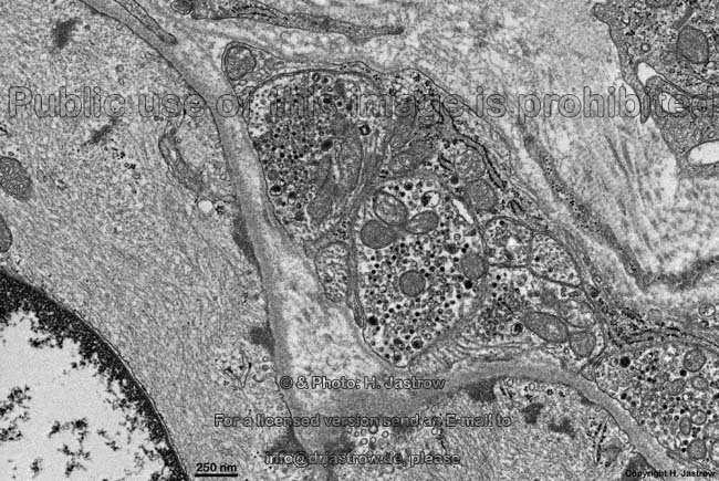
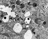
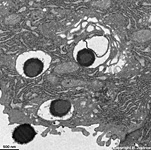
mucous und serous secretion
vesicles sublingual gland (rat) |
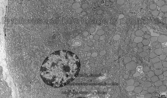
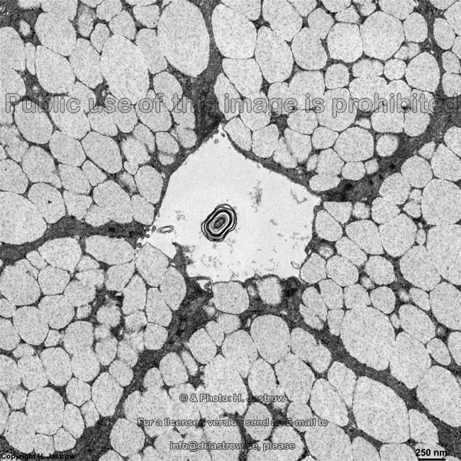
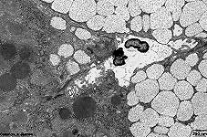
mucous epithelial cell
Glandula sublingualis (rat) |
detail: mucous
vesicles (rat) |
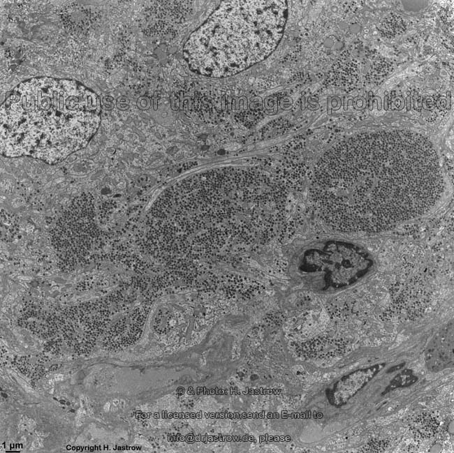
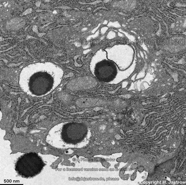
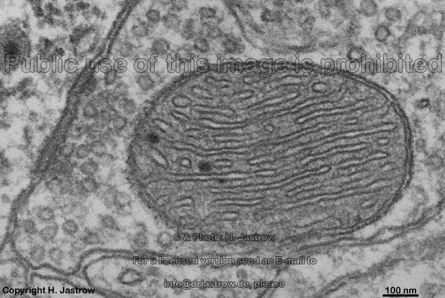
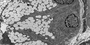
Secretion vesicles se-
minal gland (rat) |
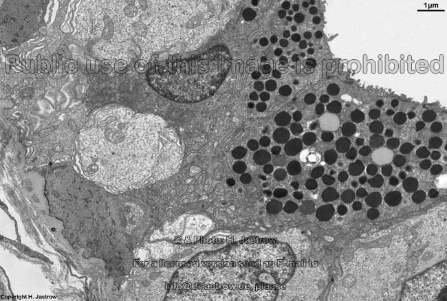
serous acinus of a small
human eccrine suet gland |
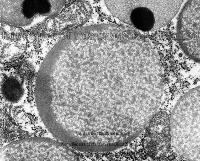
Secretory vesicles (Terminologia histologica:
Vesiculae
secretoriae) are vesicles bordered by a unit
membrane that deliver secretory products to the extracellular
space which in many cases is a glandular
acinus or the lumen of an excretory duct. The wrong, but established term
secretory granule should not be used since true granules like glycogen
granules, in contrast to secretory vesicles never have a limiting membrane.
Secretory vesicles may derive from the Golgi
apparatus (Golgi vesicles) or from rough
or smooth endoplasmic reticulum or in
some cases directly from endocytotic or modified endocytotic
vesicles as e.g., in membrane vesicles of the crusta of the ureter. They
are transported to the cell membrane,
their membrane fuses with the cell
membrane whereby the latter diverges at the point of adhesion and
the content of the vesicle is released. This process is termed
exocytosis.
Exocytotic vesicles in contrast to common endocytotic
vesicles have no clathrin coat. The size of secretory vesicles may
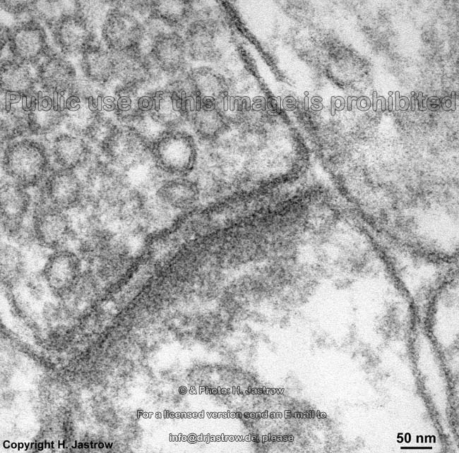
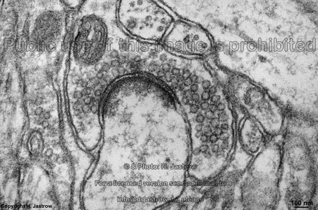
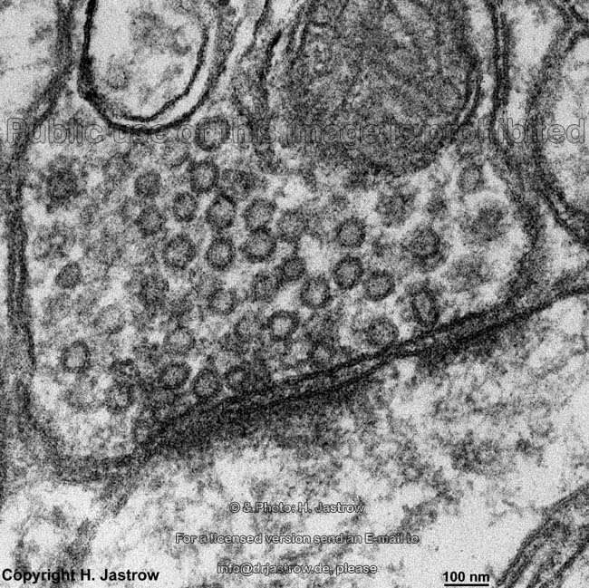
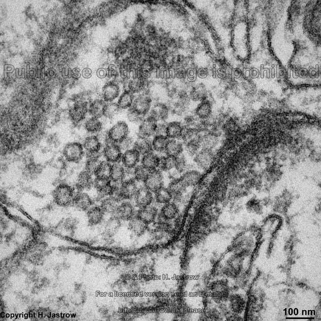
vary considerably neurotransmitter
vesicles that serve in signal transduction processes on chemical synapses
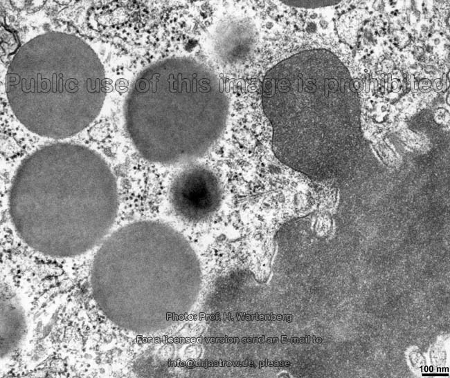
have a mean diameter of 50 nanometers (nm). Secretory vesicles of
vesicular secreting endocrine gland cells,
i.e. cells secreting directly into blood
or lymph vessels usually are about 100 nm
in diameter. Such secretions are called hormones (messenger substances
delivered to their targets i.e. receptors via the blood). The secretory
vesicles of exocrine gland cells that deliver their content into
gland
acini or excretory ducts are quite different in size in some cases over
1 µm and vary in electron-density depending on the content. This
content virtually always contains water, ions and often several different
soluted proteins e.g., zymogen vesicles of the exocrine epithelial
cells of the pancreas are comprised of a
mixture of different still inactive froms of enzymes. Examples of vesicluar
secreting endocrine glands, i.e. those that deliver secretion products
via vesiclular exocytosis are the pituitary,
adrenal
gland, thyroid gland (C-cells) and
the pancreas. Examples of exocrine glands
that release secretory vesicles are the pancreas,
the salivary glands, the lacrimal gland,
goblet
cells and the mammary gland.
Regarding the consistency and viscosity of the secretory products secretory
vesicles can be classified as:
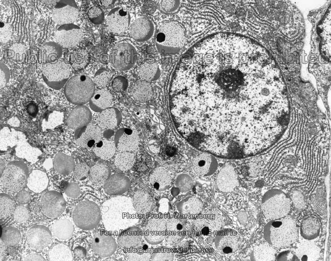
1.) serous, i.e. vesicles have a water-like
non-viscous content. Most of the proteins encountered in serous secretion
vesicles are enzymes that often are secreted in inactive preforms. Due
to the mostly relatively high protein content the vesicles are quite electron-dense
and homogenous. They mostly to not join with each other before exocytosis
and thus can be well differentiated from each other. The cytoplasm
of serous secreting gland cells is homogenous.
The cells are often seen bordering serous glandular acini. Here the cells
show their round nuclei in the basal mostly
basophilic (due to high content in ribosomes and ribosomal RNAs) cytoplasm.
Typical glands with serous secreting cells are the parotid
gland, the lacrimal gland, the pancreas
and sweat glands.
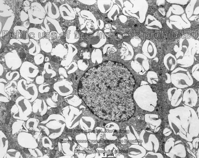
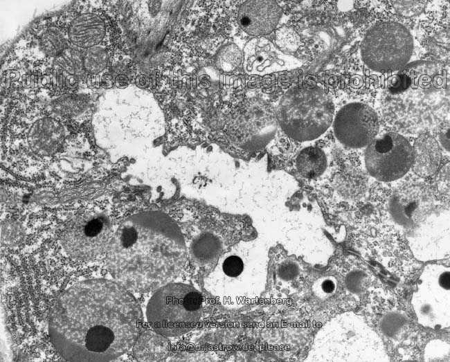
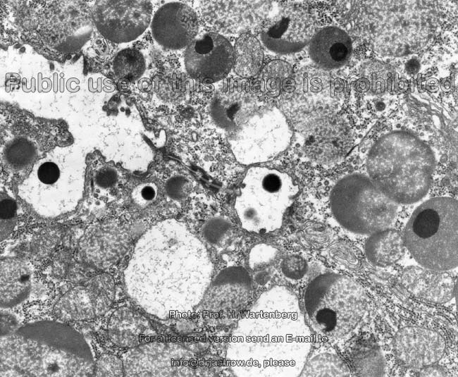
2.) mucous, i.e. highly viscous
secretion vesicles which contain acid mucins. The mucoid substances
(glycoproteins or mucopolysaccharids in most cases) can be visualized using
the PAS staining metod in light microscopy. They have not such a high affinity
to electron-microscopic staining agents therefore vesicles appear much
less electron-dense than serous ones. In many cases vesicles fuse before
being released resulting in a less electron-dense honeycombed appearance
of the apical cytoplasm. The nuclei
of mucous epithelial cells usually are flat
and located close to the basis of the soma.
Mucous secretory vesicles are present in epithelial cells of the stomach,
in goblet cells, in the
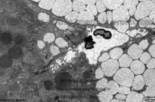
sublingual-
and submandibular gland. The last mentioned
glands also have populations of serous epithelial cells that may be present
either in acini alone or in form of vonEbner's
half moons beyond mucous epithelial cells. In these cases the serous
cells do not have direct contact to the lumen thus the intercellular clefts
between the mucosus cell are dilated to allow the serous secretions to
reach the lumen of the gland. Von Ebner's half-moons are only seen in the
sublingual-
and submandibular gland.
The delivery of secretions may happen in three different ways
of which the eccrine is most common:
1.) eccrine
= merocrine secretion, i.e. exocytosis, vesciles dock
on and fuse with the cell membrane and
deliver their content into the extracellular space without any notable
loss of cell volume since no cytoplasm
gets lost. The secretion product is water-like and non viscous in most
cases. Eccrine secretion is typical for: salivary
glands, pancreas and sweat
glands
2.) apocrine secretion, i.e. apocytosis,
"secretory vesicles" that are lipid droplets
gather in the apical part of the cell close to the lumen, integral membrane
proteins of the cell membrane like Butyrophilin
bind to proteins associated to the border of the lipid
droplets thereby the latter begins to protrude into the lumen. By further
binding to the base of the spherical lipid droplets
the adjacent cell membrane fuses and
the lipid droplet, the surrounding small seam
of cytoplasm and the covering cell
membrane are released. Thus some cell
membrane and a little cytoplasm gets
lost with every secretion process resulting in a small reduction of the
cellular volume. The fat droplet is set free, when later on the surrounding
membrane collapses. This rare kind of secretion is only seen in the mammary
gland, apocrine sweat gland cells and the glands of Moll in the eyelid.
3.) holocrine
secretion, i.e. holocytosis, the whole gland cell fills up with
more and more "secretory vesicles" that in fact are lipid
droplets that may fuse. Thereby the epithelial
cell looses its contact to the basement
membrane and is pushed forward towards the lumen by proliferation of
the basal cell layer. The farer the cell gets to the basement
membrane the worse the situation for its nutrition by diffusion, thus
the cell begins to degenreate. The nucleus and
the cell organelles dissolve and the fatty,
sebaceous secretion product fills the whole cell. When contact to neighbouring
cells gets lost by desmosome disintegration,
the whole cell becomes the secretory product in that the cell
membrane finally disrupts and the rest of the cytoplasm
as well as the lipid droplets are released.
This type of secretion only occurs in sebaceous
glands and the glands of Meibohm or Zeiss of the eyelid.
4.) avesicular secretion, The
majority of glands shows secretory vesicles,
however, in case the chemical properties of the secretion product (size,
lipophily) do not require concentration and packing in vesicles, secretory
vesicles are not produced. A lot of substances are secreted directly from
the cytoplasm of exo- or/and endocrine
cells via more or less specific transmembrane
proteins. This is not visible in the electron microscope. The endocrine
pineal
gland is an example for an avesicular secreting gland. Its lipophilic
melatonin can easily pass the cell membrane
without need of any transporter. The liver
is an exocrine gland that secrets gall without any vesicles into bile
canaliculi and at the same time releases hundrets of other substances
via Disse's space into the blood, i.e. it is
exo- and endocine, in case one uses the expression endocrine not only for
hormones. In eccrine secretion, which also is avesicular, transport
proteins in the cell membrane secrete invisily ions which then is followed
by paracellular flow of extracellular fluid.
An English page with detailed information and more images is only available
in the professional version of this atlas.
--> glands in general, salivary
glands, synapse
--> Electron microscopic atlas Overview
--> Homepage of the workshop
Some images were kindly provided by Prof. H. Wartenberg;
other images, page & copyright H. Jastrow.