Overview adrenal gland (Glandula
suprarenalis):
Pages with explanations are linked to the
text below the images if available! (Labelling is in German)
 |
 |
 |
 |
 |
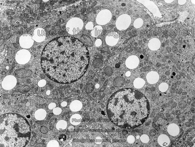
| Zona reticularis 1 (monkey) |
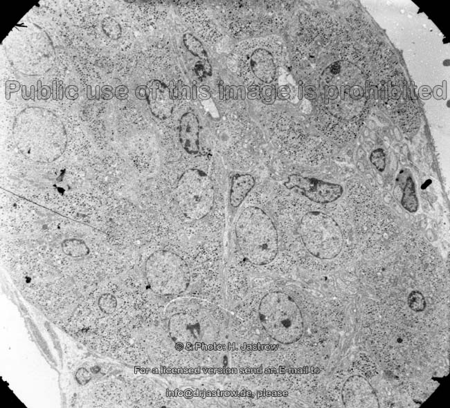
Zona reticularis 2 (monkey) |
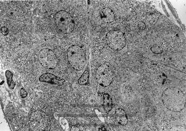
Zona reticularis 3 (monkey) |
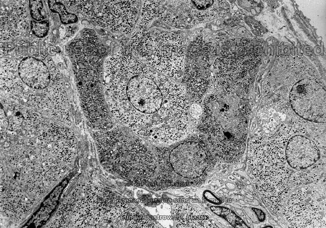
Zona reticularis 4 (monkey) |
Zona reticularis 5 (monkey) |
The adrenal gland (suprarenal gland; Terminologia histologica:
Glandula suprarenalis) is an endocrine
gland and has a weight of about 10g. It consists of an outer zone with
high endocrine activity called adrenal cortex and an inner zone the adrenal
medulla.The adrenal gland is covered by a capsule
(Terminologia histologica: Capsula) consisting of an outer layer of tight
connective
tissue (Terminologia histologica: Lamina fibrosa; englisch: fibrous
lamina) and a deeper cellular lamina (subcapsular blastema; Terminologia
histologica: Lamina cellulosa) with small cells of low differentiation.
The capsule is surrounded by the
loose
connective tissue of the renal adipose
capsule which is rich in blood vessels and
fat
cells.
Adrenal cortex (Terminologia
histologica: Cortex)
Different layers of the cortex can be distinguished:
1. Zona glomerulosa (Terminologia
histologica: Zona glomerulosa corticis). This outermost layer comprises
about 10% of the cortical thickness and lies directly beyond the capsule.
The endocrine epithelial cells located here have a little less lipid droplets
and produce mineral corticoids, mainly
aldosteron,
which is a part of the renin-angiotensin-system. In consequence the cells
are controlled by feedback-regulation by means of angiotensin II. The following
2. Zona fasciculata (Terminologia
histologica: Zona fasciculata) is much stronger and makes up about 70 %
of the mass of the cortex. The Zona fasciculata is mainly producing glucocorticoids
like Cortisol und Cortison but also releases few amounts of estrogenes
and androgenes. It is controlled by the adrenocorticotropic hormone of
the pituitary. The next layer is the
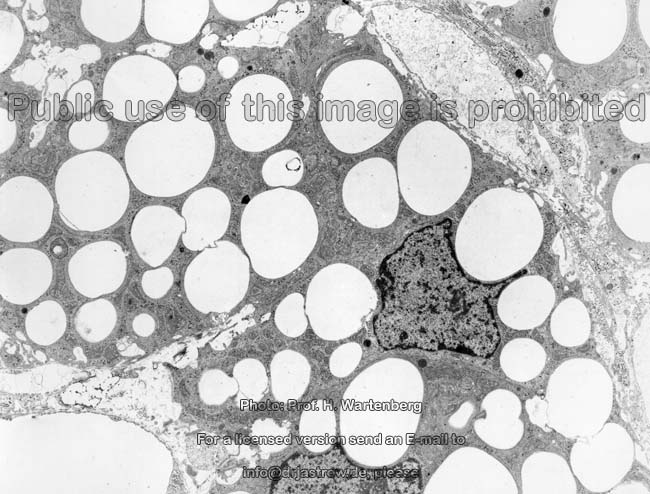
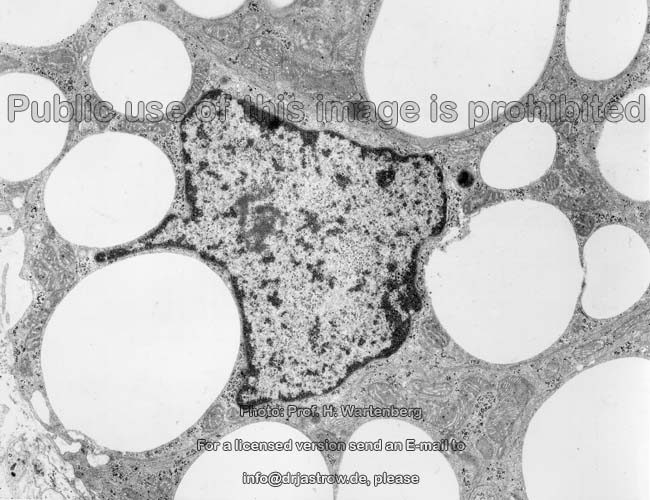
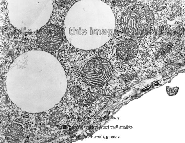
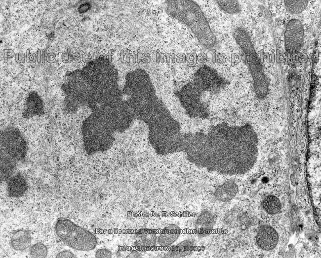
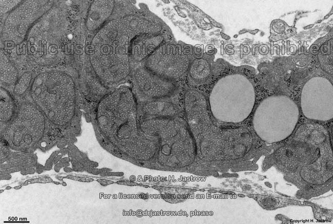
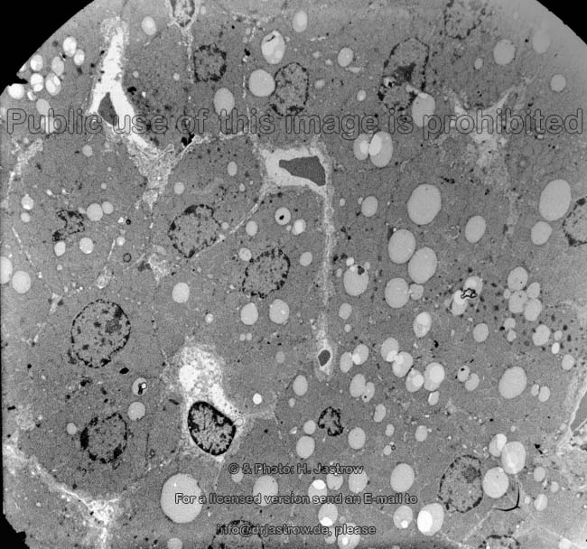
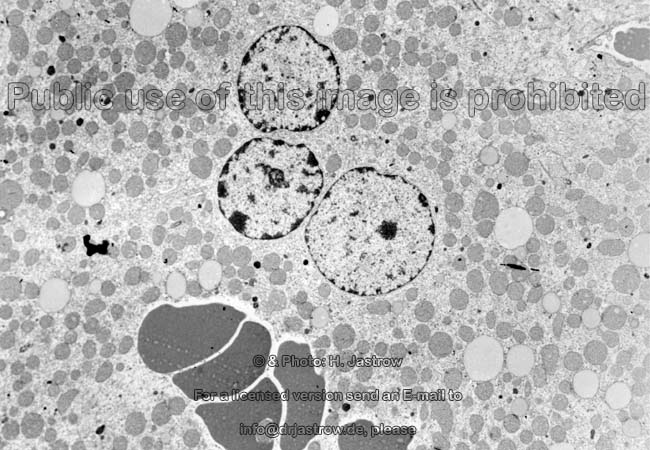
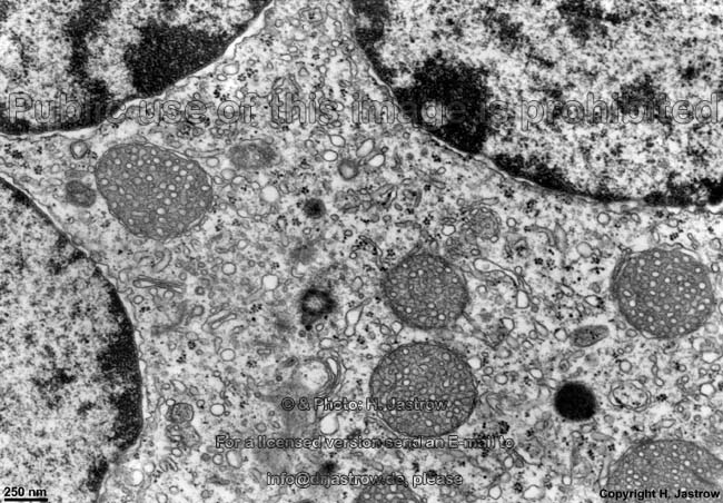
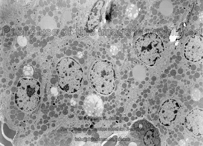
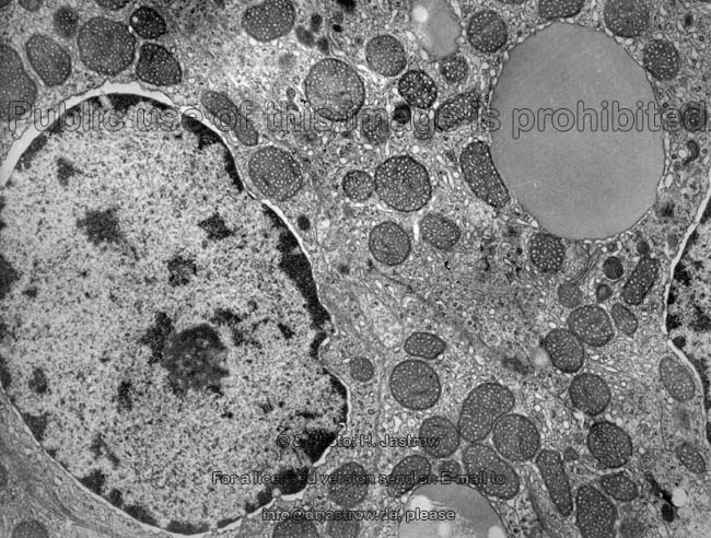
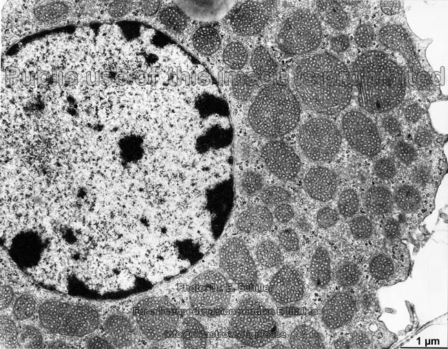
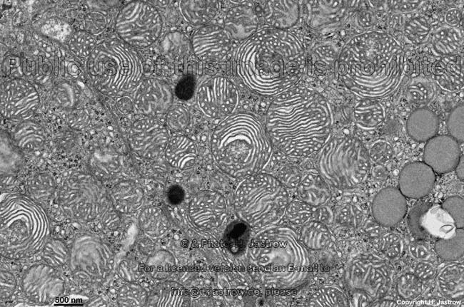
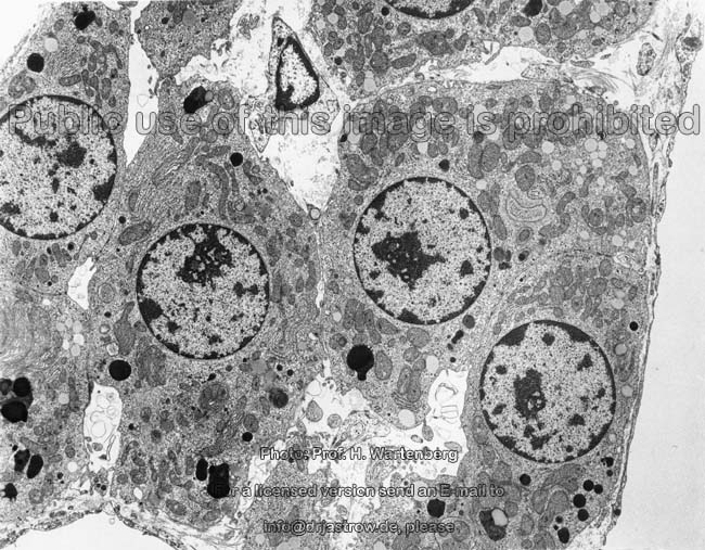
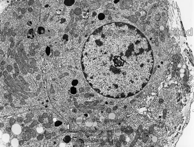
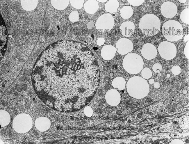
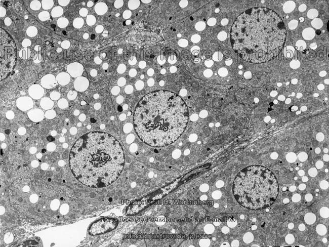
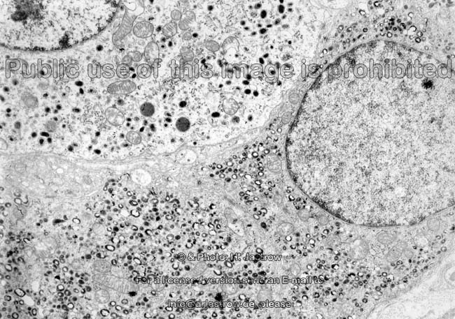
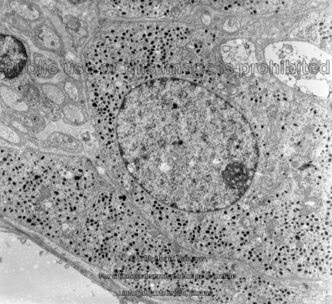
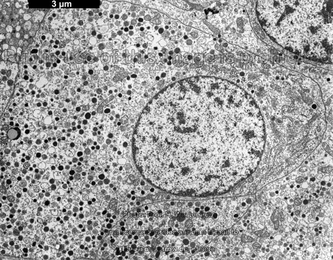
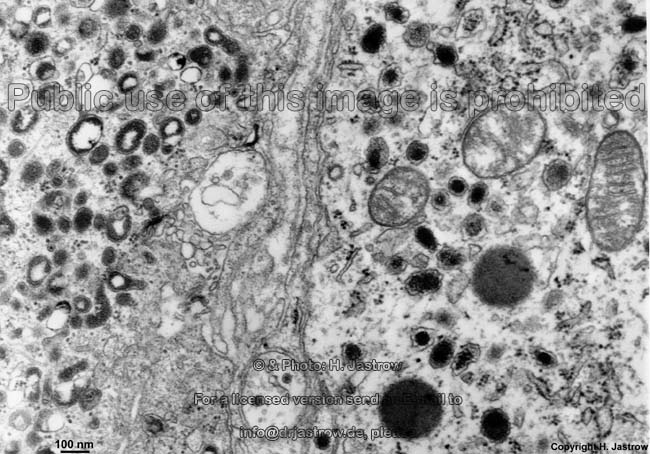
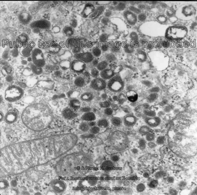
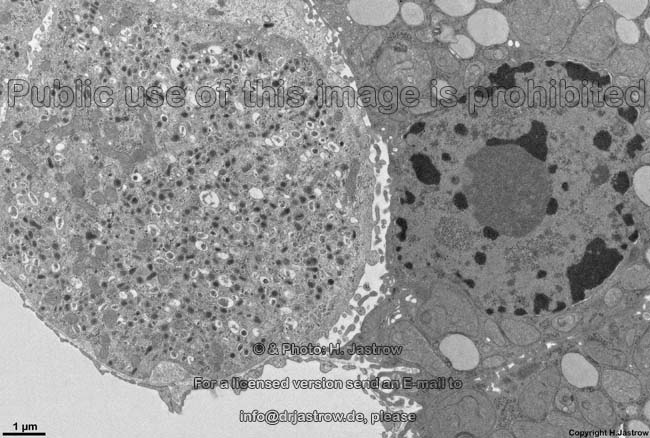
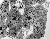
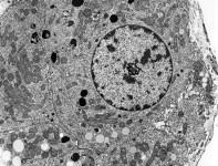
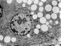
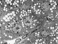
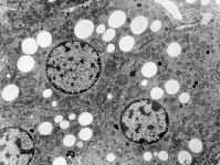
3. Zona reticularis (Terminologia
histologica: Zona reticularis). This zone consists of a loose network of
capillaries
and cell cords and comprises about 20% of the mass of the cortex. The zona
reticularis releases
androgenes which are not very effective and
are further processed in other tissues like prostate,
mamma or
placenta to gain more efficiency.
While in males testosteron from the interstitial
cells of the testis is the main androgen,
adrenal androgenes are those of relevance in females.
Adrenal medulla (Terminologia
histologica: Medulla)
The adrenal medulla is in fact a ganglion of the sympathicus with large
multipolar autonomic ganglion cells
of the sympaticus (large cells, "true neurons", Terminologia histologica:
Neurona multipolaria autonomica) are seen intermingled with endocrine cells,
i.e. medullary chromaffin cells (Terminologia histologica: Endocrinocyti
medullares).
The adrenal medulla releases adrenalin
and noradrenalin in high rates under
stressy conditions as well as some neuropeptides.
More detailed information and images are available in the professional
version of this atlas.
--> glands, kidney,
pituitary,
thyroid
gland, ganglia,
mitochondria
--> Electron microscopic atlas Overview
--> Homepage of the workshop
Some images were kindly provided by Prof. H. Wartenberg
or Dr. E. Schiller; other images, page & copyright H. Jastrow.