Overview kidney (Ren):
Pages with explanations are linked to the
text below the images if available! (Labelling is in German)
|
|
|
|
|
|
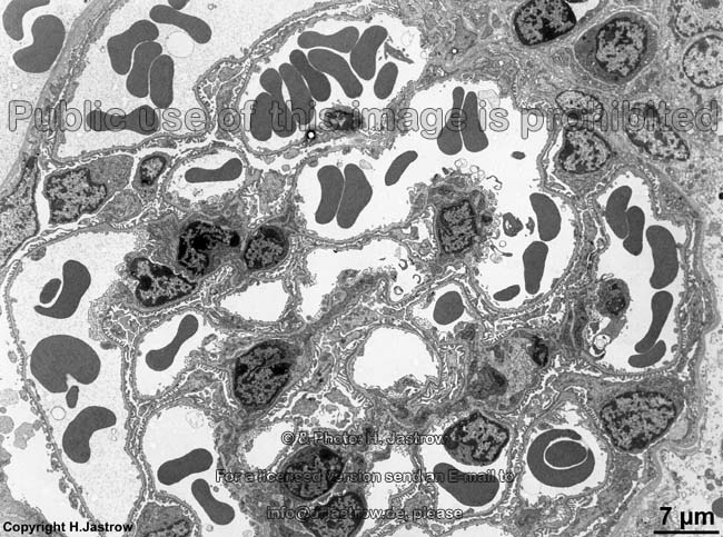
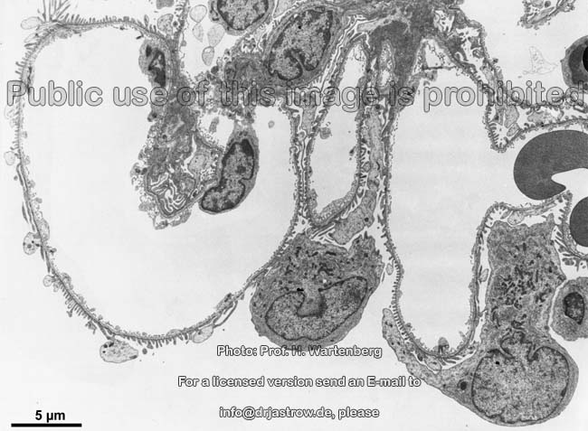
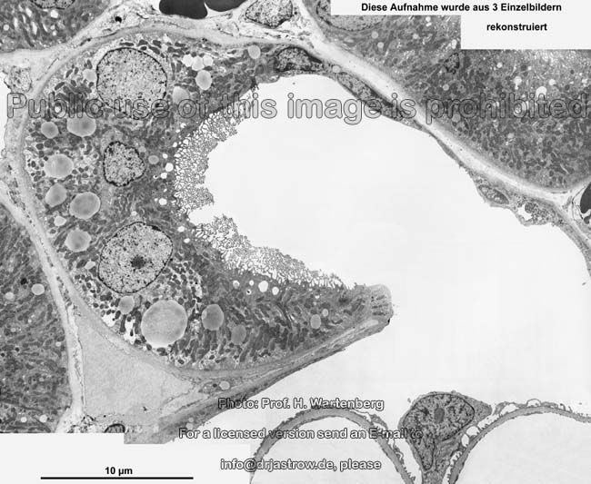
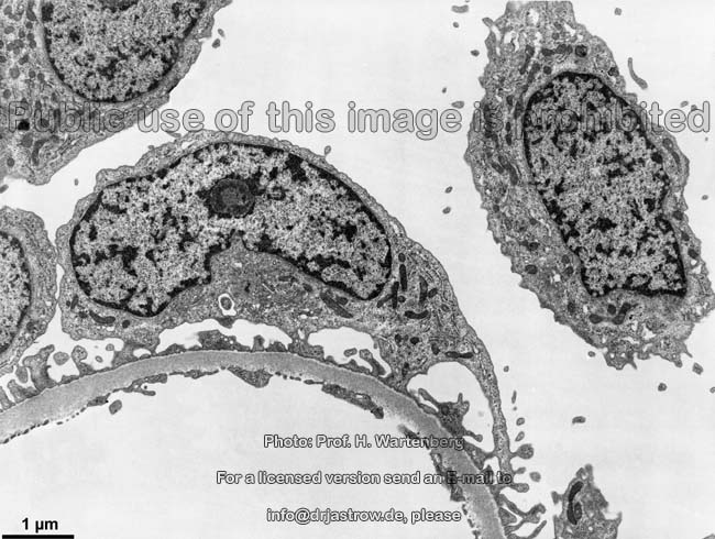
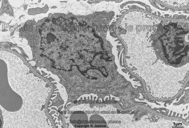
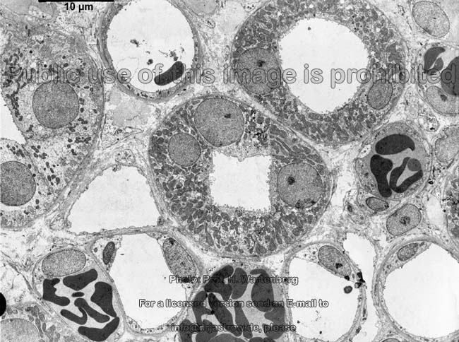
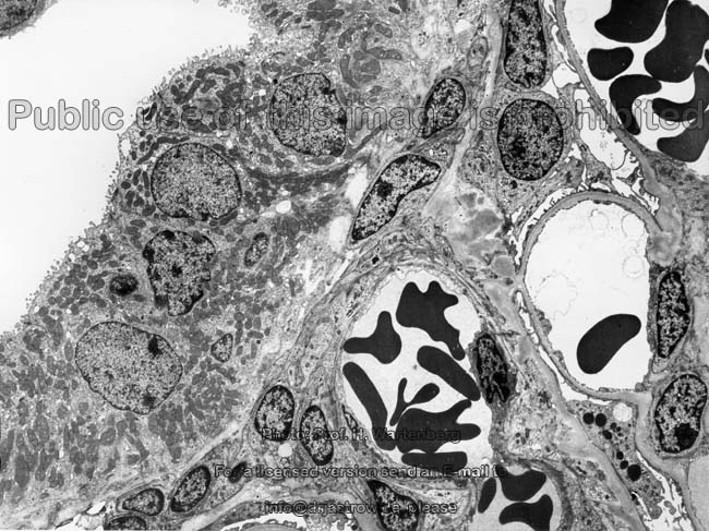
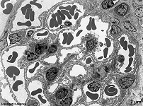
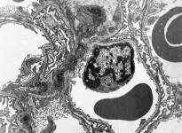
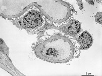
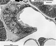
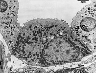
glomerulum
overview (rat) |
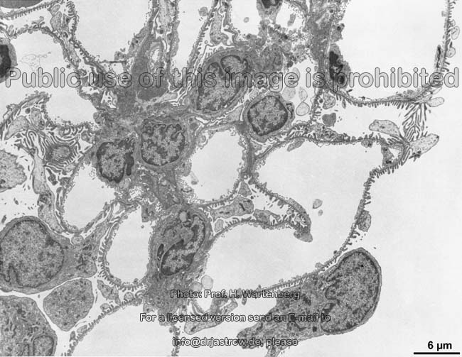
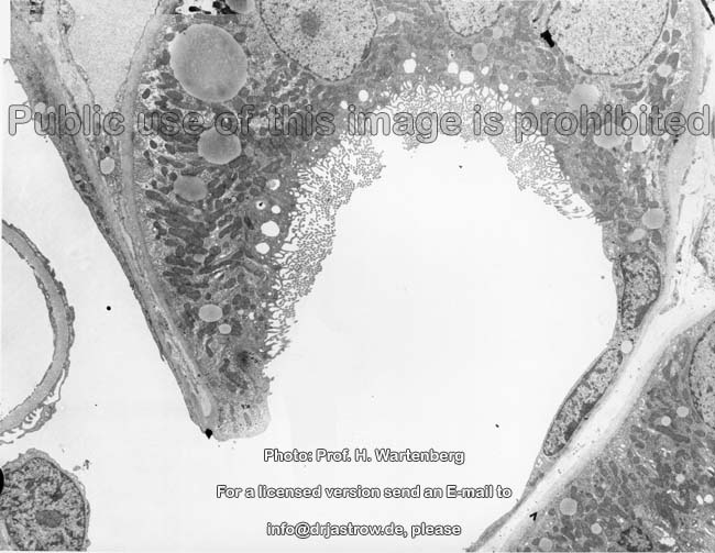
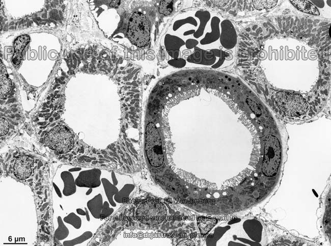
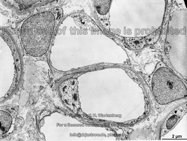
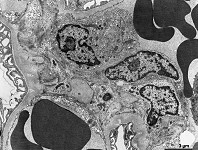
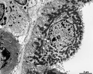
glomerular capillary
loops (monkey) |
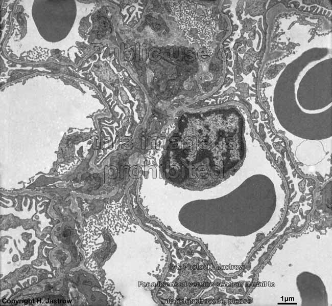
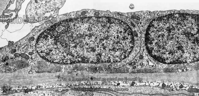
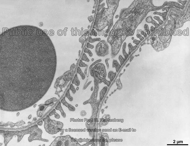
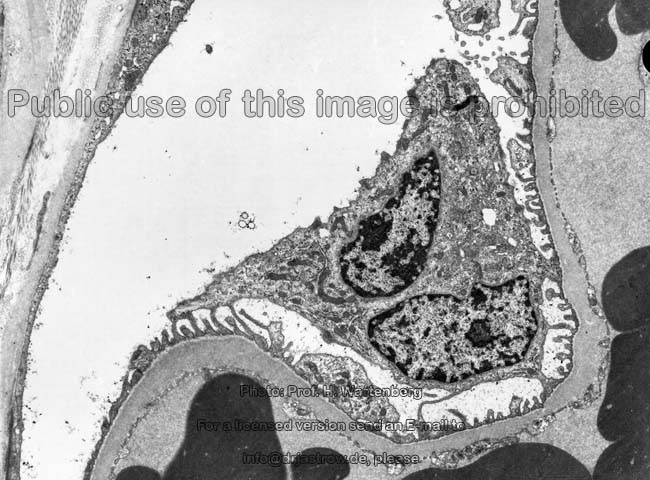
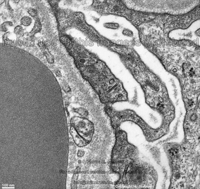
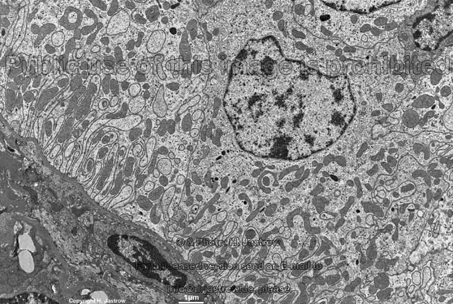
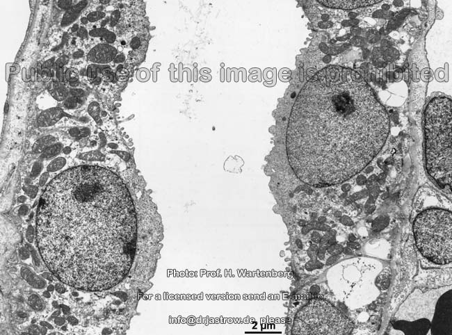
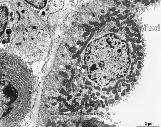
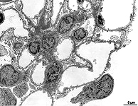
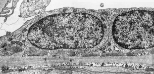
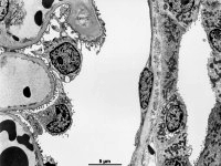
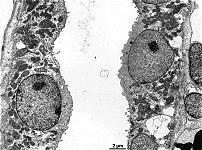
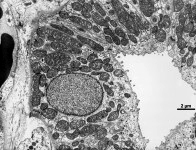
endothelial cell of a
glomurular capillary (rat) |
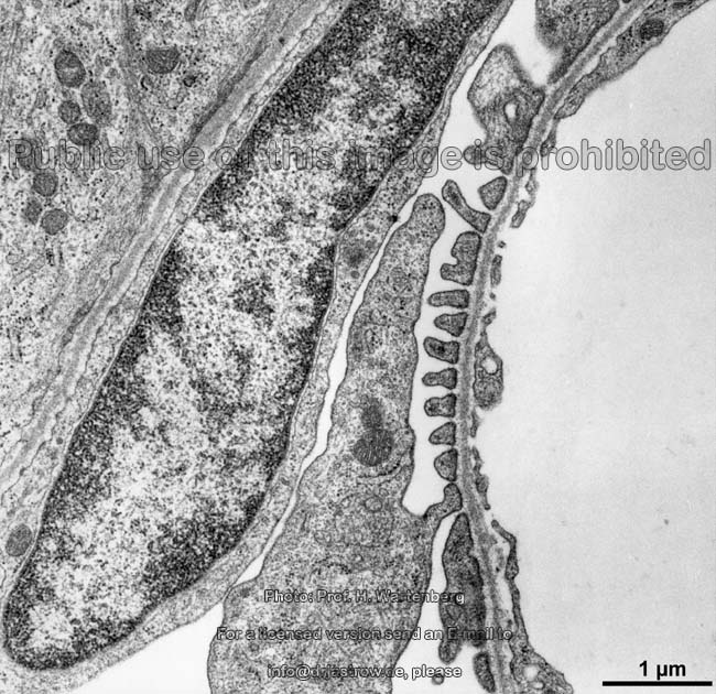
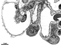
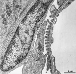
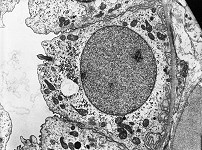
glomerulum detail
(monkey) |
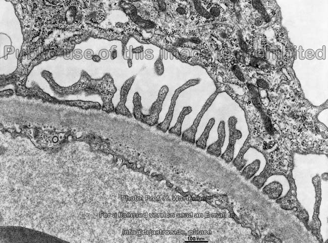
glomerulum detail 2
(monkey) |
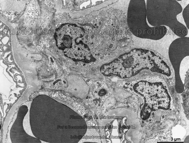
glomerulum detail 3
(monkey) |
|
|
|
|
|
|
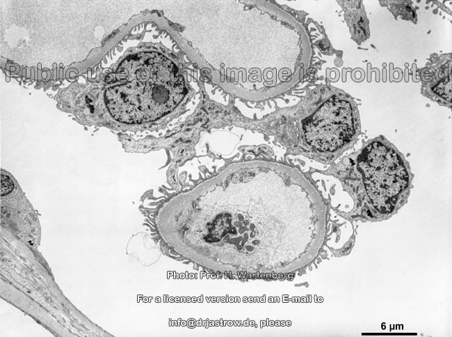
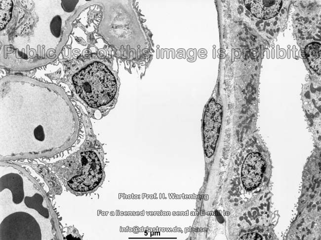
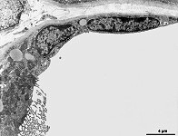
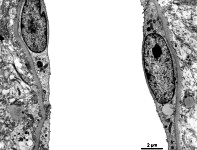
cells of the parietal part of
Bowmann's capsule (monkey) |
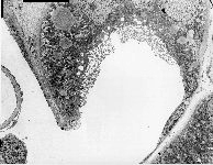
Bowmann's capsule
+ glomerulum (monkey) |
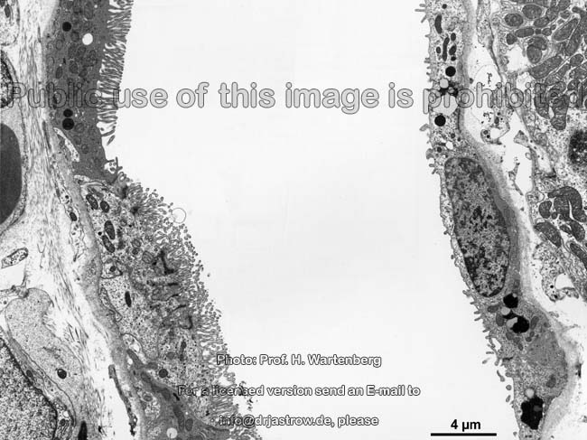
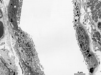
urinalry pole change
of epithelium (monkey) |
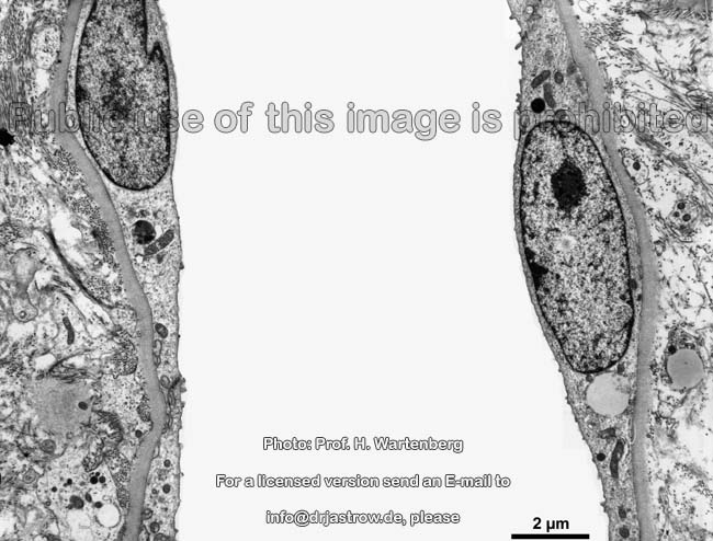
urinary pole 1
(monkey) |
urinary pole 2
(monkey) |
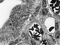
glomerulum and
distal tubule (monkey) |
 |
 |
 |
 |
 |
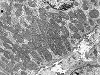 |
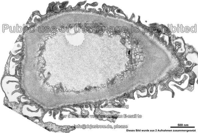
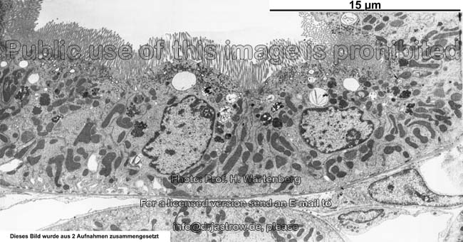
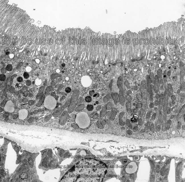
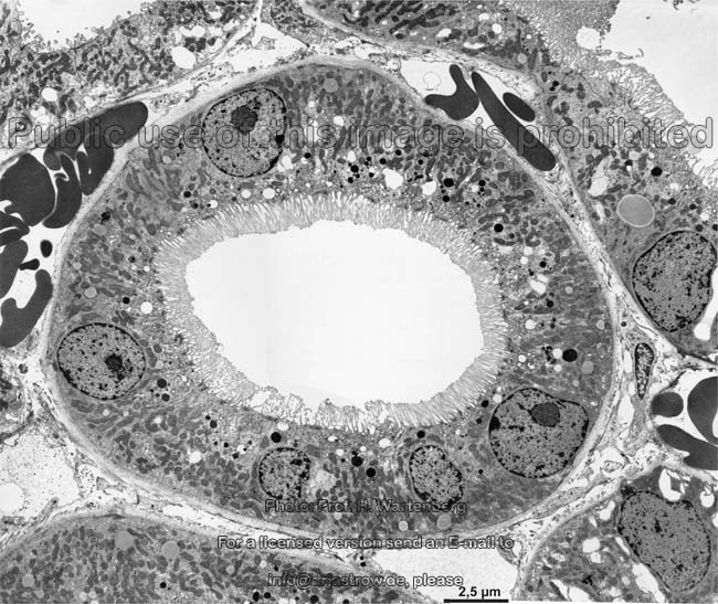
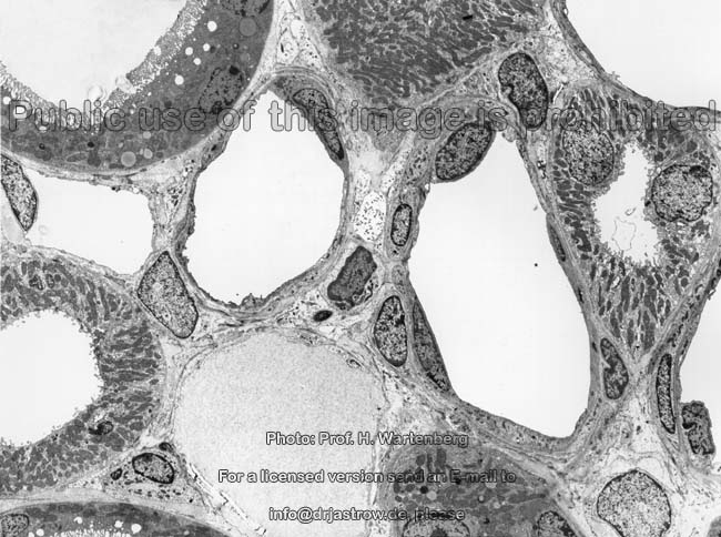
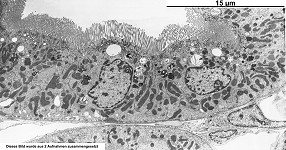
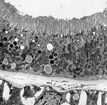
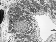
mikrovilli on epithelial cells of the
proximal renal tubule (monkey) |
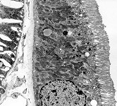
proximal
tubule 2 (monkey) |
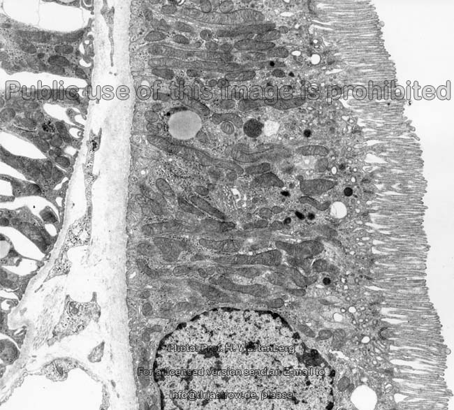
proximal renal tubule
3 (monkey) |
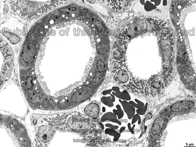
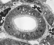
proximal renal tubule in
coss-section (monkey) |
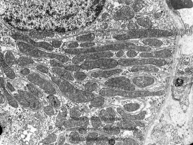
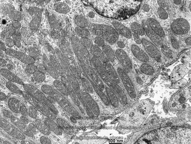
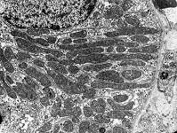
basale striping of aproxi-
mal tubule cell (monkey) |
basal striping 2
proximal tubule (monkey) |
|
|
|
|
|
|
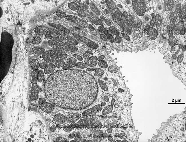
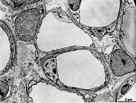
Henle's loop in cross-
section (monkey) |
Henle's loop in longi-
tudinal section 1 (monkey) |
Henle's loop in longi-
tudinal section 2 (monkey) |
distal convolut 1
(monkey) |
distal renal tubule 1
(monkey) |
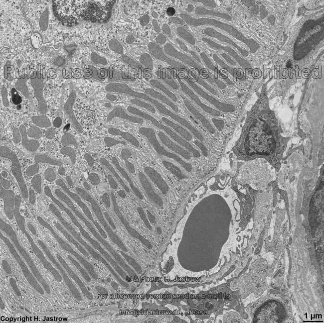
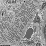
basal stripes of mito-
chondria (rat) |
|
|
|
|
|
distal tubule + blood
vessels (monkey) |
distal convolut 2
(monkey) |
distal convolut 3
(monkey) |
distal renal tubule 2
(monkey) |
collecting duct
(monkey) |
The kidney (Terminologia histologica: Ren, Nephros) has
a weight of 120 - 200 g and serves in filtration of urine from blood.
Thus it is the initial organ of the urinary system (Terminologia
histologica: Systema urinarium) that also comprises the draining ureter
which reaches the bladder (Vesica urinaria)
and then continues into the urethra.
Overview:
The kidney is covered and protected by a perirenal
fat capsule (perinephric fat; Terminologia histologica: Capsula adiposa)
consisting of a usually some centimetres wide construction fat of unilocular
fat tissue cells. However, at the beginning, i.e. in newborns and babies
and then dramatically decreasing in number multilocular
fat cells are present here as well. The organ proper is ensheated
by a fibrous capsule (Terminologia
histologica: Capsula fibrosa) build of woven
connective tissue. One human kidney has 6 to 9 lobes (Renculi;
Terminologia histologica: Lobi renales) which usually are not visible as
it is the case in many animals (e.g., cows). Human kidneys with a clearly
visible lobulation are called renculus kidneys and present in ~ 7% of adults.
During development humans have 14 single kidney premordia thus a maximum
of 14 lobes is possible for each side. The lobes can be recognised in kidney
sections by their calyces into which they release
their urine. Renal columns (Bertini columns; Terminologia histologica:
Columnae renales) are present in the regions where the lobes border each
other they reach downwards from the renal cortex
and are in fact parts of the renal cortex which to the exterior borders
the renal capsule. In the other direction
the renal cortex borders the renal
medulla which ends in the calyces that
collect the urine and lead it into the renal pelvis.
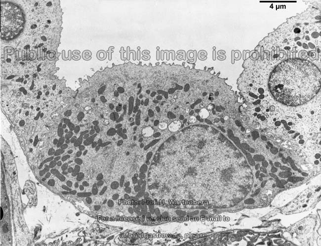
The functional unit of the kidney is the Nephron,
which consists of the renal corpuscles
and the non branched segments of the tubules,
that together with 11 other such tubules terminate in one collecting
duct. About 150 to 180 Litres of primary urine are filtered
from blood in the renal
corpuscles located only in the
renal cortex.
This urine flows through initial proximal tubules
that reach down into the medulla then make a
turn in Henle's loop ascend as distal
tubules pass a chloride ion sensor, the macula
and continue to connecting tubules turn
downwards again and via collecting ducts descend
to papillary ducts ending at the tip of the
papilla
of the medulla. Here the final urine is released
into the renal calices. While passing the nephron
about 99% of the water and lots of ions are regained from the primary urine.
Further, other substances are secreted here. This results in 1.5 Litres
of
final urine. These processes happen either transcellularly,
i.e. through the
epithelial cells with help
of transport proteins in apical and basal cell membranes or,
restricted to some sections of the tubules, paracellularly (for
small ions and substances) through the pores
of the
tight junctions of the junctional
complex between the cells via the intercellular
space. Additionally, the kidney releases the following hormones:
renin and erythropoëtin.
Detailed information and many more images are only available in the
professional version of this atlas.
--> blood-urine barrier,
ureter,
urinary
bladder, prostate,
seminal
gland, epithelium,
microvilli
--> Electron microscopic atlas Overview
--> Homepage of the workshop
Many images were kindly provided by Prof. H. Wartenberg;
other images, page & copyright H. Jastrow.