Overview capillaries (Vasa
capillaria):
Pages with explanations are linked to the
text below the images if available! (Labelling is in German)
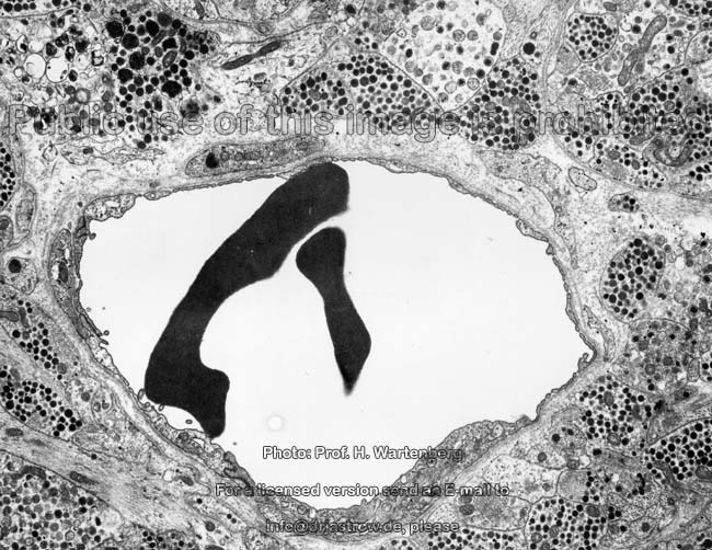
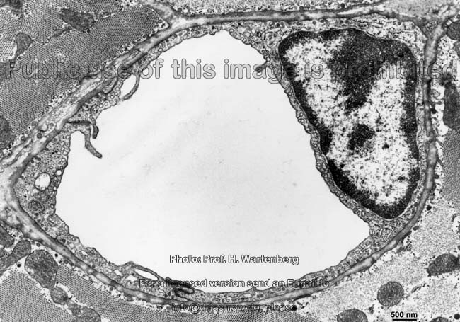
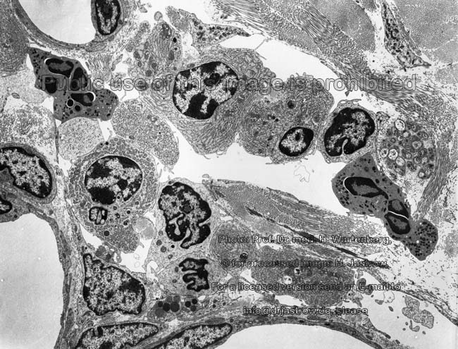
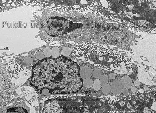
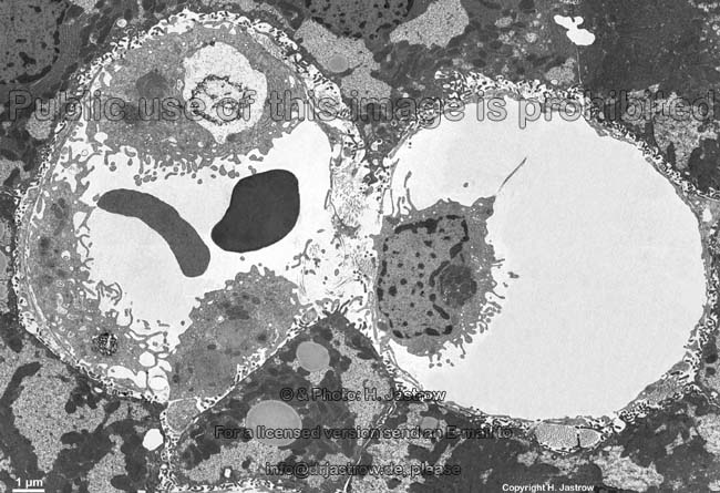
Capillary vessels or simply capillaries (Terminoligia
histologica: Vasa capillaria) are the smallest blood
vessels. Their diameters vary about that of an erythrocyte
(red blood cell; diameter ~ 7.5 µm) whereby
the smallest capillaries even require an adaptive deformation of the erythrocyte
since their lumen is only 6 µm in width. The largest capillaries,
however, have diameters of up to 15 µm. Usually a capillary
forms a capillary arch (Terminoligia
histologica: Ansa capillaris) as described here: they receive blood
from the metarteriols
which drain into arterial capillaries
(Terminoligia histologica: Vasa capillaria arterialia, Vasa precapillaria;
~
8 µm in diameter) which begin when the smooth
muscle cells and the tiny internal elastic
membrane are no longer detectable. The
following midcapillaries (Terminoligia histologica: Vasa capillaria
intermedia) are only 6-7 µm in width. Here the blood
flows with a speed of about 0.5 mm per second and the intravascular
pressure
is between 2-4 kPa (15 - 30 mm Hg). The
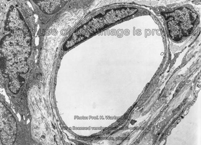
next portion of the arch are the venous capillaries (Terminoligia
histologica: Vasa capillaria venosa, vasa postcapillaria) with a width
of 8-9 µm which drain into smallest venules
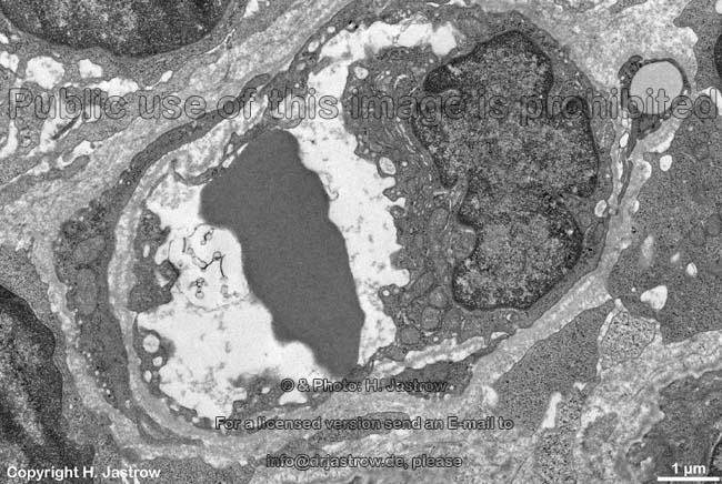
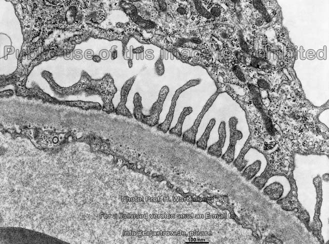
whose diameters are above 15 µm sind. In the area of the venous capillaries
and of the venules the intercellular space
between the lining flat endothelial cells partly is widened to a few hundred
nanometres which is termed (Terminoligia histologica: Apertura intercellularis,
Apertura transcellularis). This is why this is the typical location for
white blood cells to leave the vascular system to become free connective
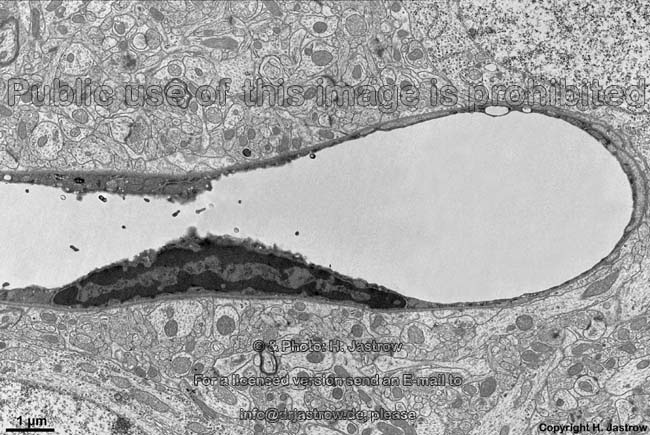
tissue cells. A very intense exchange
of metabolites is performed in the capillary region since the blood
gases and nutrients can easily reach the surrounding tissue due to the
very thin in some places additionally fenestrated wall of the capillaries
(fenestrated endothelial cells).
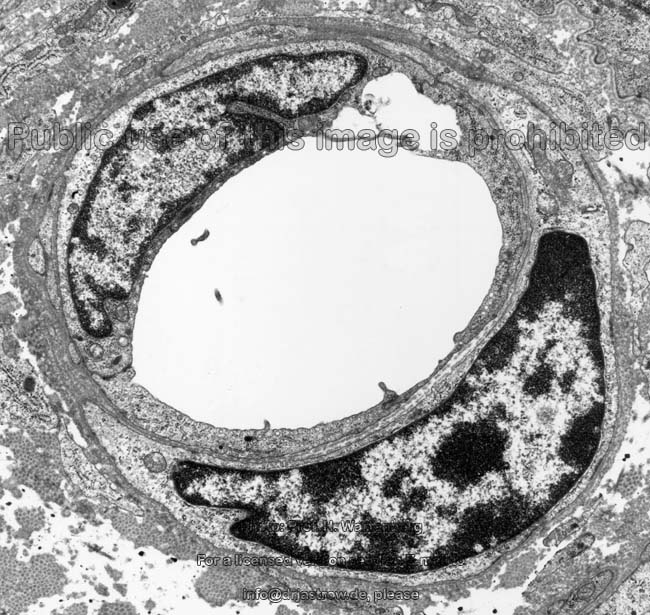
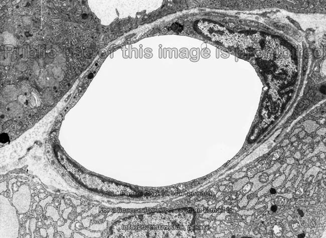
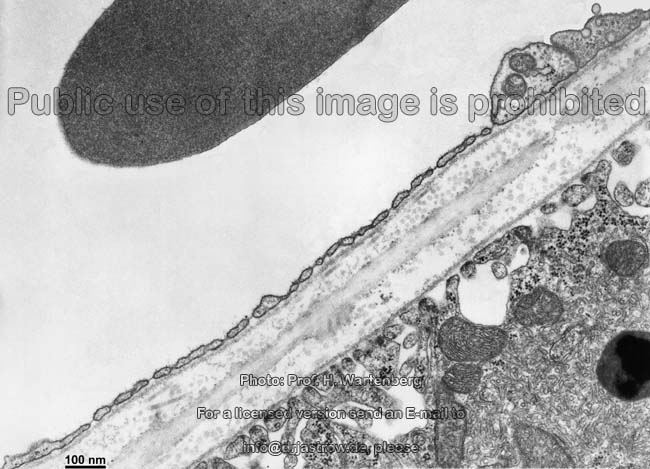
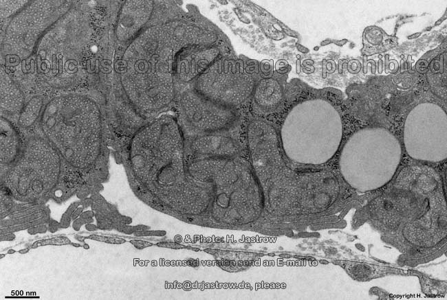
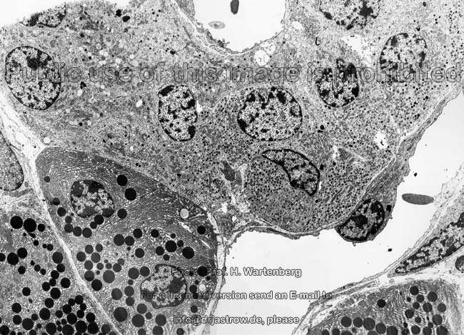
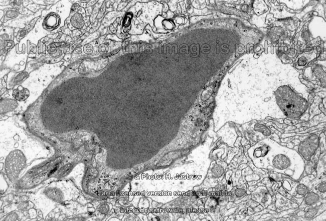
Morphology:
When looking from iside to outside a capillary
begins with an intima (missing in Terminoligia histologica; proposal:
Tunica intima, Tunica interna) consisting of only one very flat fenestrated
endothelial cell followed by a basal
membrane. Few reticular fibres (collagen
type 3) anchor the capillary to the surrounding connective
tissue. A media is missing. The
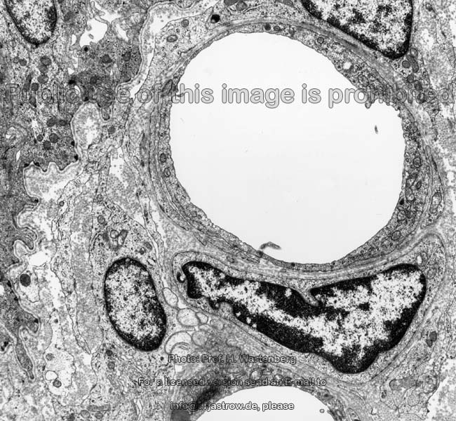
directly following adventitia (missing in Terminoligia histologica;
proposal: Tunica adventitia, Tunica externa) is variabel often
incontinuous adventitial cells (Terminoligia histologica: Cellulae
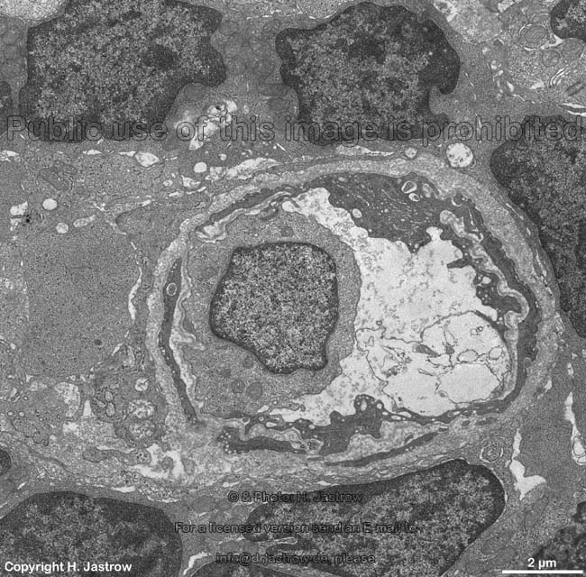
adventitiales) are present. These immotile
connective tissue cells are called pericytes (Terminoligia histologica:
Pericyti). From point of view of function and morphology they are in between
fibroblasts
and smooth muscle cells. By contraction of
the microfilaments of their cytoskeleton
(actin filaments) they can moderately influence
the width of the capillary. Depending on organ and function the chemical
composition of the basal lamina
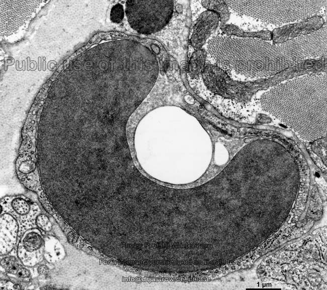
of capillaries may vary. In general we can differentiate
1. capillaries of the muscle type
with continuous endothelium
(non fenestrated endothelium; Terminoligia histologica: Endothelium non
fenestratum, Endothelium continuum) on a non-interrupted basal
lamina,
2. capillaries of the visceral
type on a continuous basal
lamina with endothelial cells showing pores covered by tiny diaphragms
forming a fenestrated endothelium
(Terminoligia histologica: Endothelium fenestratum).
--> endothelial cells,
blood
barriers, blood cells, blood
vessels, arteriole, venole
--> Electron microscopic atlas Overview
--> Homepage of the workshop
Some images were kindly provided by Prof. H. Wartenberg;
other images, page & copyright H. Jastrow.