List of abbreviations
Vocabulary
of micros-
copic
anatomy
specialist terms
explained in
English +
German

Every attempt was made to provide correct information and labelling, however any liability for eventual errors or incompleteness is rejected!

dieser Seite

Editor:
Dr. med.
H. Jastrow


Conditions
of use
Overview collagen fibres (Fibrae collagenosae):
Pages with explanations are linked to the text below the images if available! (Labelling is in German)
collagen fibrils type1
tendon (rat)
collagen fibrils with
67 nm striping (rat)
collagen fibrils
ovary stroma (rat)
collagen fibrils type 1
and 3 (rat)
type 1 + 3 collagen fibrils of sub-
cutaneous connective tissue (rat)
human collagen fibres formed
of type 1 + 3 collagen fibrils
collagen fibrils type 1 +
elastic fibres (monkey)
bundle of type 1 collagen
fibrils of monkey cornea
collagen fibres, mast cell
Ligamentum ovarii (rat)
human collagen fibrils type 1
human collagen fibrils
type 1, cross-section
detail thereof human collagen
fibrils
collagen and elastic fibres
from liver stroma (rat)
collagen fibres next to a large elastic
fibre in human subcutis

Collagen fibres (Terminologia histologica: Fibrae collageni) are stable fibres of the extracellular connective tissue consisting of collagen fibrils (Terminologia histologica: Fibrilla collageni). They may associate to each other forming larger collagen fascicles (Terminologia histologica: Fasciculi collageni). Collagen fibres are hardly fractioning light when regarded with a light microscope, however, with a polarisation microscope a compound fraction is apparent. The unbranched fibres are very resistant to traction and about 5% stretchable. They are soluble in cooking water whereby they become glue. Their Latin name derives from this fact (Kolla = glue). They swell up when acids are added, are soluble in leach and are rapidly digested by pepsin. Applying light microscopic staining procedures the have the following colours. red (haematoxylin-eosin, see image 1; "van Gieson"); blue (Azan), see image 2; green ("Masson-Goldner"), see image 3; yellow to gray-green (iron-haematoxylin). In the electron microscope longitudinally cut collagen fibrils are some micrometers long and show characteristic bands (see below). In cross-sections the fibrils have round profiles with diameters of 50 to maximal 200 nm.
The table shows 14 different types of collagen (for further ones cf. special literature):
type chains composition of triple helix triple helix length, structural details occurrence, specialities
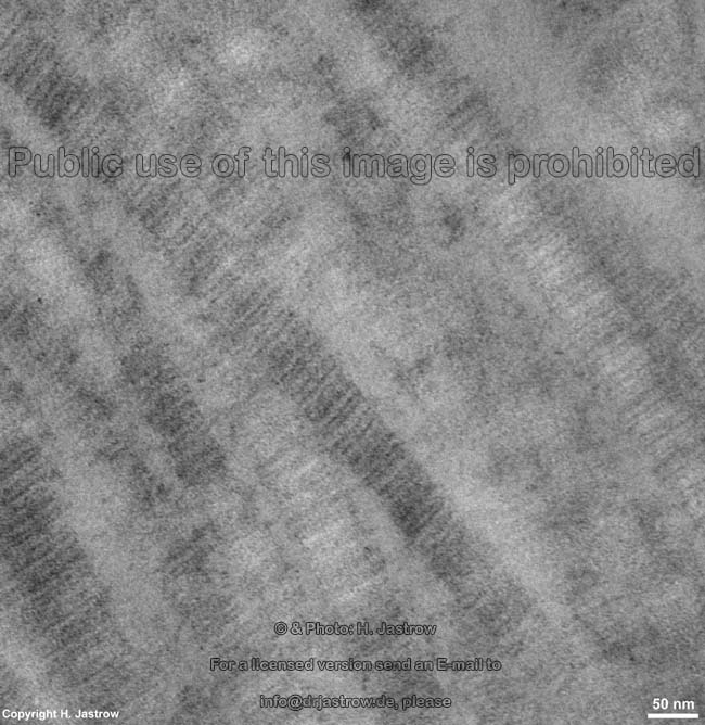
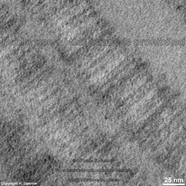
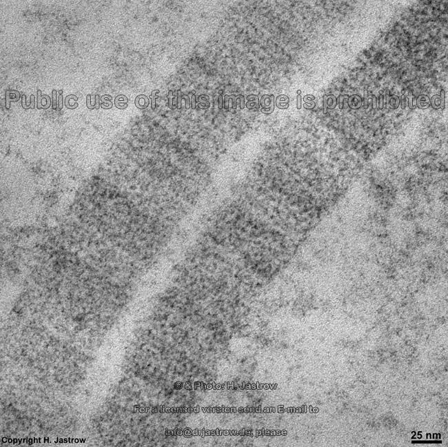
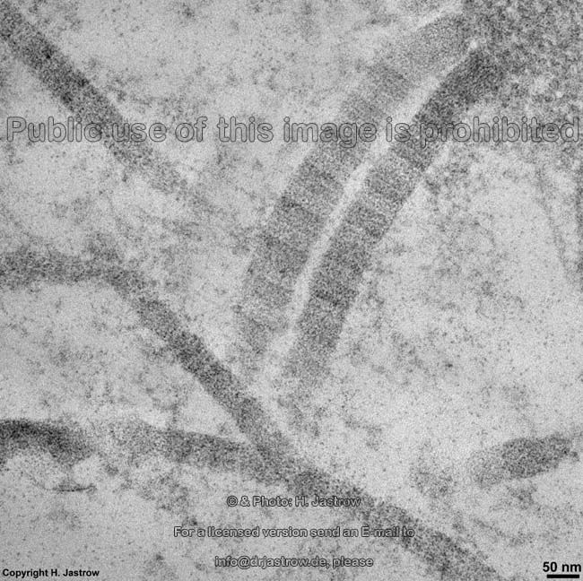
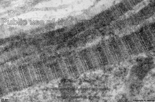
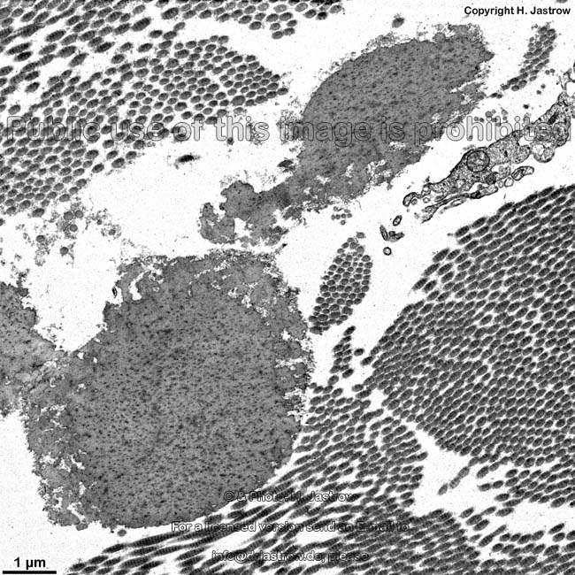
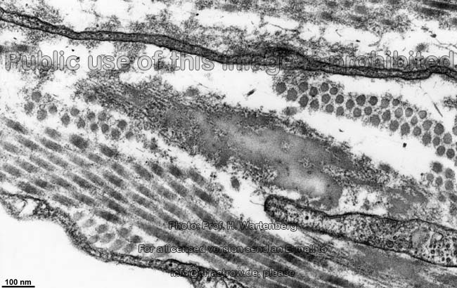
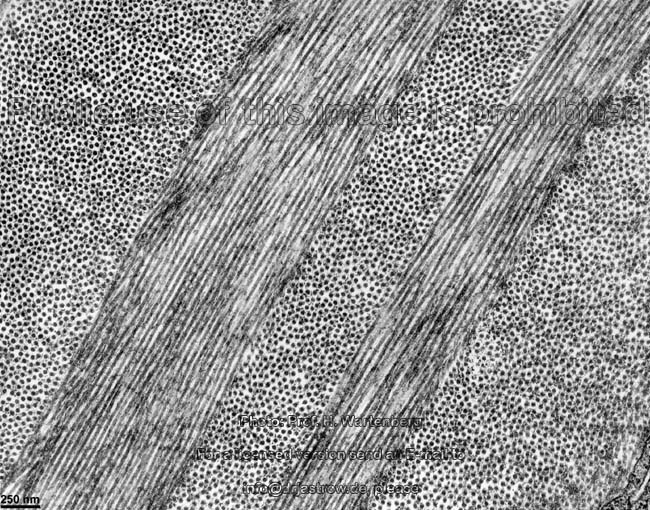
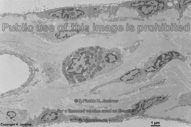
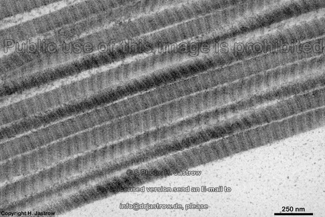
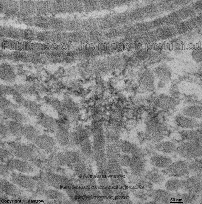
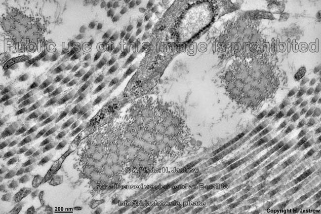
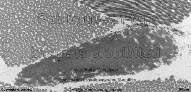
collagens with long fibrils with 67 nm periodicity of bands fibrils may consist of different types of collagens, e.g. type I + III + V or type II + XI
I
a1(I); a2(I) [a1(I)]2[a2(I)] 300 nm; diameter of fibrils 50 to maximal 200 nm
chains 1.050 amino acids long 
subcutis, tendon, bones, dentin, Bowman's membrane, sclera & cornea of the eye
in nearly all kinds of connective tissues but not in cartilage
If
a1(I); a2(I) [a1(I)]3 300 nm foetal form
II
a1(II) [a1(II)]3 300 nm; diameter of only ~30 nm;
covered by a viscous matrix of proteoglycans
hyaline- and fibrous cartilage, vitreal body of the eye,
chorda dorsalis, tectorial membrane of the inner ear
III
a1(III) [a1(III)]3 300 nm; diameter of ~40 nm;
usually together with type I
in type I fibrils, form reticular fibres, loose connective tissue, corneal stroma,
subcutis, muscles, walls of blood vessels
V
a1(V); a2(V); a3(V) [a1(V)]3, [a1(V)]2[a2(V)]
und [a1(V)][a2(V)][a3(V)]
390 nm; N-terminal globular domain
usually together with type I
in type I fibrils, foetal tissues and membranes; interstitial tissue, bones, Bowman's membrane, tectorial membrane, around smooth muscle cells, corium, placenta
XI
a1(XI); a2(XI); a3(XI) [a1(XI)][a2(XI)][a3(XI)] 300 nm; often together with type II, thin fibres in type I fibrils, hyaline cartilage, tectorial membrane
fibril associated collagens with a triple helix which is disrupted by globular domains
IX
a1(IX); a2(IX); a3(IX) [a1(IX)][a2(IX)][a3(IX)] 200 nm; N-terminal globular domain;
covalently bound to the surface of type II fibrils
cartilage, vitreous body, tectorial membrane, binds glycosaminoglycans
XII
a1(XII) [a1(XII)]3 large N-terminal globular domain; cross-like
molecule, bound to the surface of collagen type I
embryonal tendons, subcutis, corneal stroma
XIV
a1(XIV) [a1(XIV)]3 large N-terminal globular domain;
cross-like molecule
fetal subcutis, tendons
fibril associated collagens which form string of beat-like filaments
VI
a1(VI); a2(VI); a3(VI) [a1(VI)][a2(VI)][a3(VI)] 150 nm; N- and C-terminal globular domain; bands with
periodicity of 150 nm, collagen type I associated
in most interstitial tissues, at the connection of muscles to tendons,
pericellular matrix of cartilage, in the walls of blood vessels, in the endomysium
collagens that show leaf-like association
IV
a1 to 5(IV) [a1(IV)]2[a2(V)]
and other forms
60 nm long triple helix region and 40 nm long
globular domains; formation of tetramers 
all basement membranes, formation of a two-dimensional interconnected network,
is secreted by cells of endothelium, epithelium, glia cells and fat cells
VIII
a1(VIII); a2(VIII) ? regular triangular gutter subendothelial tissue, Descemet's membrane of the cornea of the eye
X
a1(X) [a1(X)]3 150 nm; C-terminal globular domain  secreted by hypertrophic chondrocytes thus present in the
epiphyseal plate during growth of bones
collagens which are anchoring fibrils
VII
a1(VII) [a1(VII)]3 450 nm; dimer; globular domain on both ends forms the short anchoring fibrils which connect the basal lamina of epithelia to
the deeper loose connective tissue whenever a basement membrane is present
collagens of which only the DNA was shown
XIII
a1(XIII) ? ? only the c-DNA of this protein was shown in endothelial cells
The most common types of collagen are I, II and III. When longitudinally cut the collagen types I, II, III, V and XI show a characteristic periodicity of 67 nm, which is due to a regular shift of the single molecules in the fibril. Biochemically the proteins of which the collagens consist have a-chains with Gly-Pro-X as most common sequence of amino acids, whereby X can be any amino acid.
The synthesis of collagens takes place on the ribosomes of the rough endoplasmic reticulum in resident connective tissue cells, mostly fibroblasts or fibrocytes. Signalling peptides are linked on the primarily single chains, then single proline and lysine rests are hydroxylated and oligosaccharids are coupled to them. Further, galactose is bound to hydroxylysine. The chains aggregate to a first triplehelix and make disulfide bridges. The triple helical procollagen is formed from the C to the N terminus and then is transported to the Golgi apparatus where glucose is linked to the O-coupled oligosaccharids. Then the complex is transported via Golgi vesicles to the cell membrane for exocytosis. After release into the extracellular space N- and C-terminal propeptides are uncoupled resulting in the tropocollagen molecule which is the unit component of the fibrils. The latter is 1.2 nm thick and has a length of 300 nm. The single tropocollagen molecules aggregate in a way that they are shifted by one quarter of their length from lateral to make covalent binding to each other. Thus less than 18 nm thick filaments are formed which are not yet striated. The latter grow slowly by further aggregation of tropocollagen to form the final striated collagen fibrils. Collagen fibres then are formed by aggregation of a considerable number of such fibrils which then are interlinked by aggregation of proteoglycans. Collagen fibres are of different sizes depending on the kind of tissue and local biomechanical requirements and have a characteristic texture and a main direction of course.

--> other fibres in connective tissue: elastic, reticular
--> ground substance, mobile and immobile connective tissue cells, connective tissue
--> Electron microscopic atlas Overview
--> Homepage of the workshop


Two images were kindly provided by Prof. H. Wartenberg; other images, page & copyright H. Jastrow.