Overview skeletal muscle (Textus
muscularis striatus skeletalis):
Pages with explanations are linked to the
text below the images if available! (Labelling is in German)
|
|
|
|
|
|
|
|
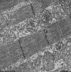
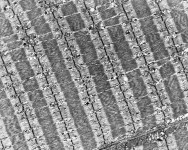
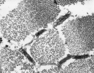
contracted human
eye muscle 1 |
contracted human
eye muscle 2 |
contracted human
eye muscle 3 |
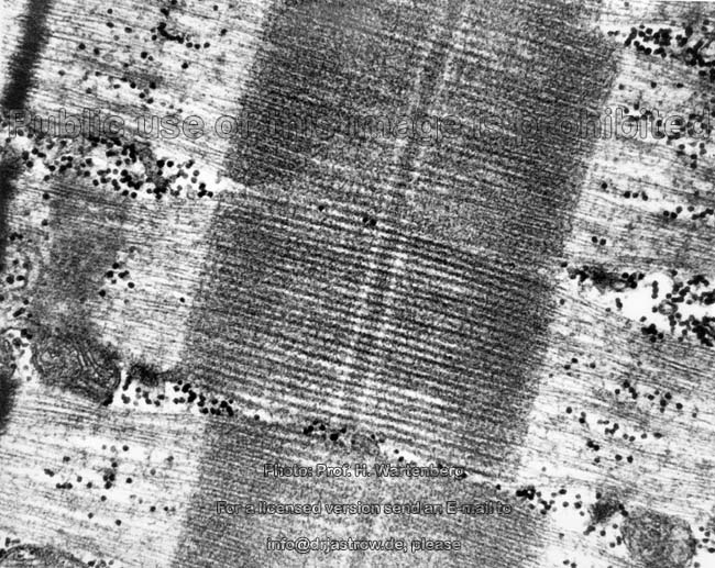
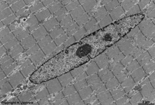
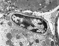
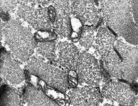
human eye muscle
overview |
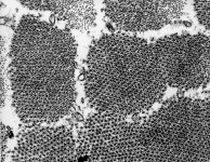
uncontracted human
eye muscle |
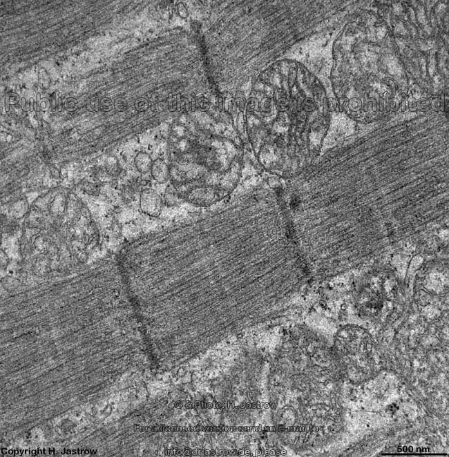
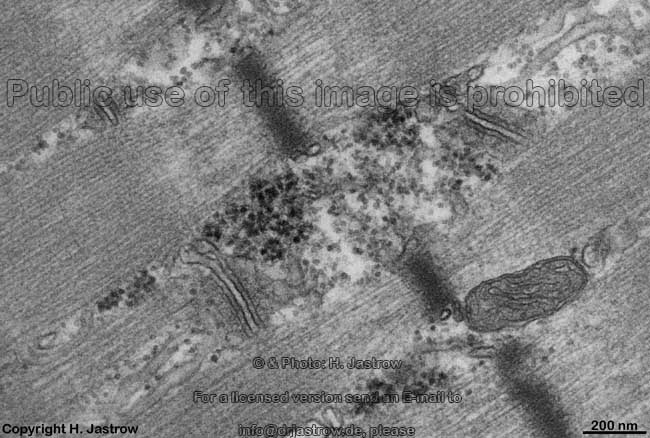
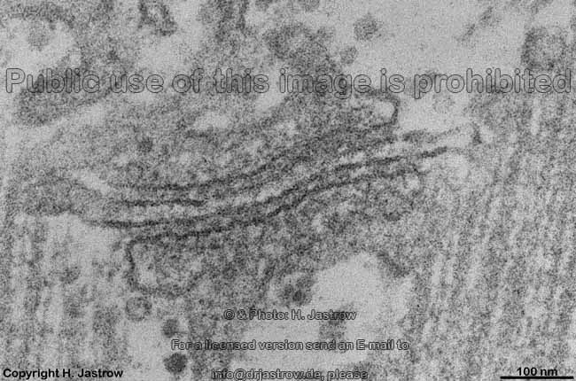
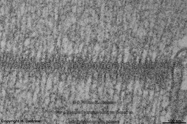
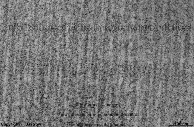
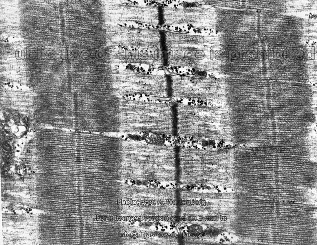
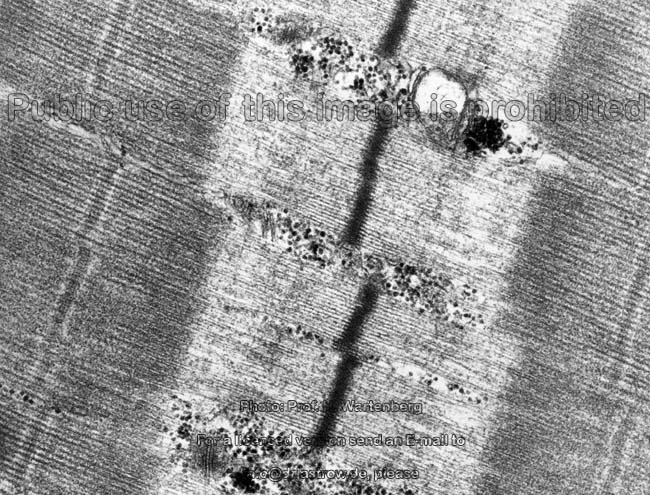
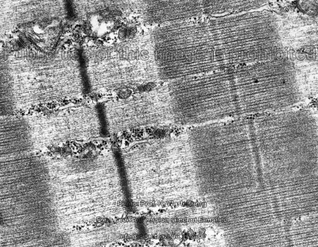
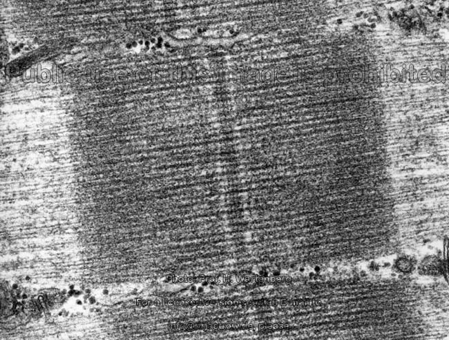
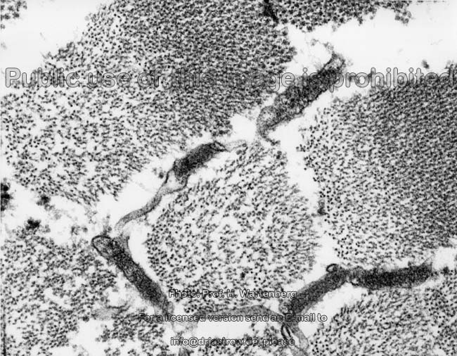
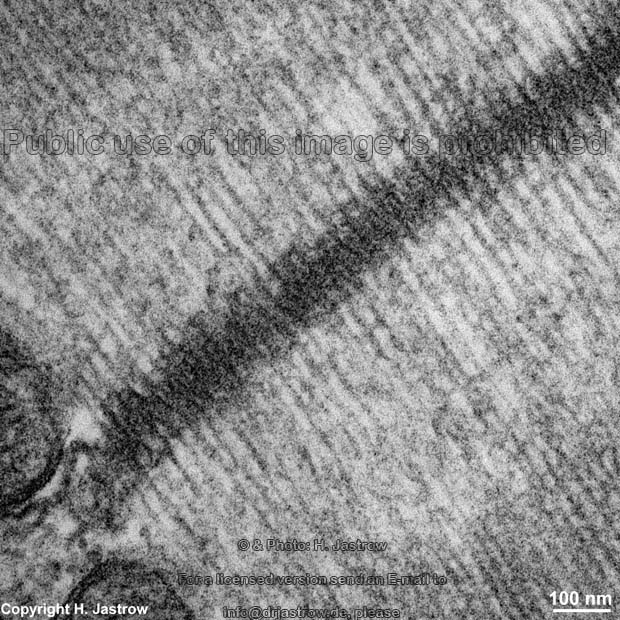
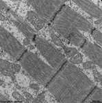
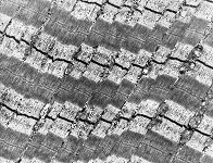
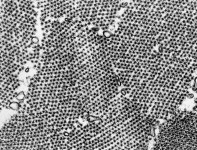
T- and L- Tubuli, beta-Glykogen
granules 1 (rat) |
T- + L- Tubuli, beta-Gly-
kogen granules 2 (rat) |
T- and L- Tubuli 3
(rat) |
 |
|
|
|
|
|
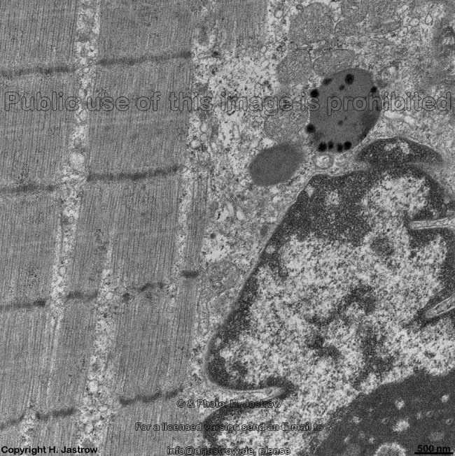
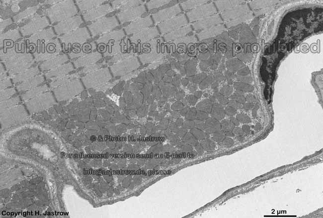
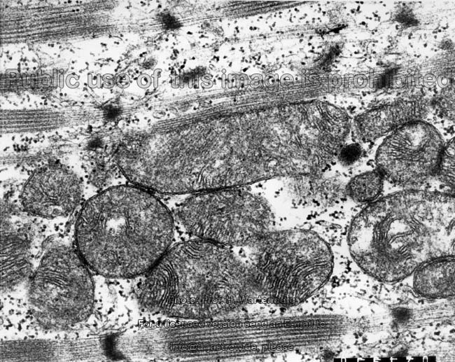
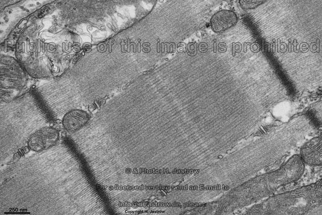
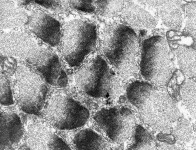
very large mitochondrium
(rat) |
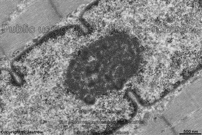
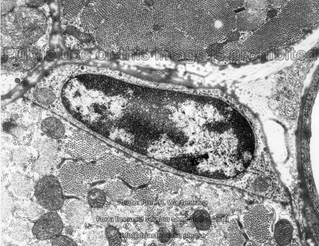
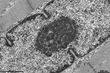
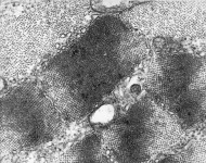
nucleus and nucleolus
(rat) |
detail thereof |
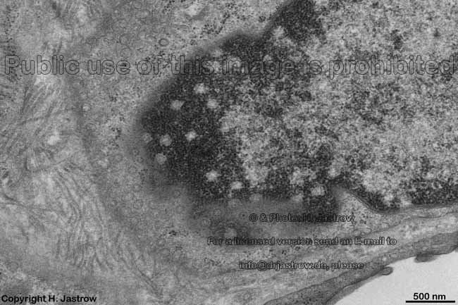
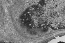
nuclear pores myocyte
(rat) |
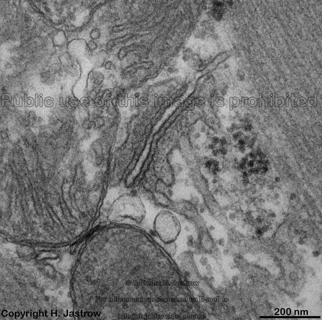
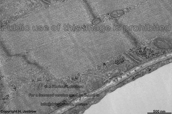
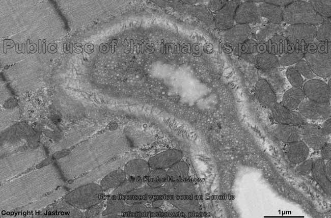
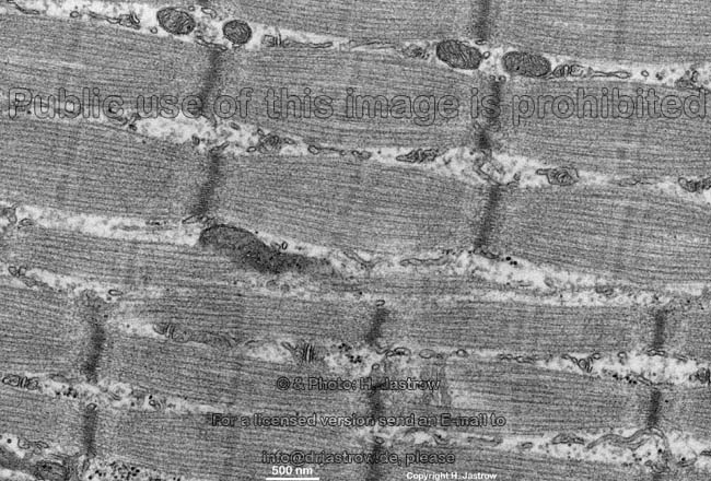
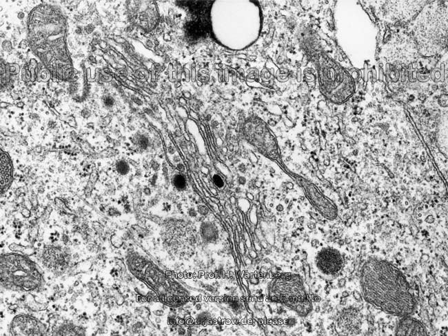
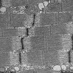
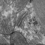
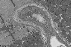
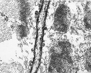
T-tubule, endothelium,
capillary (rat) |
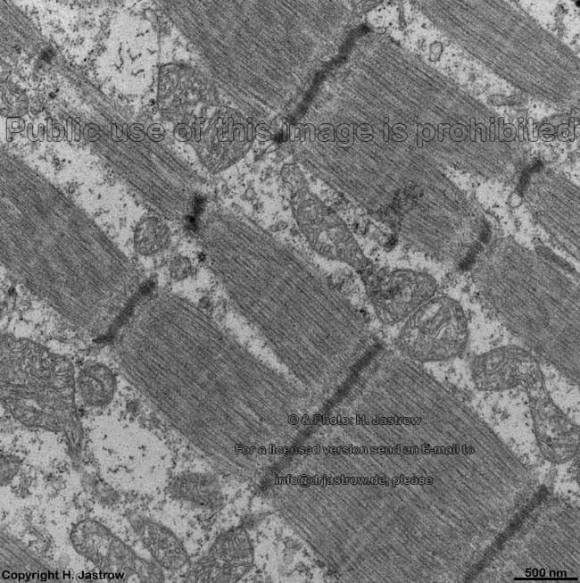
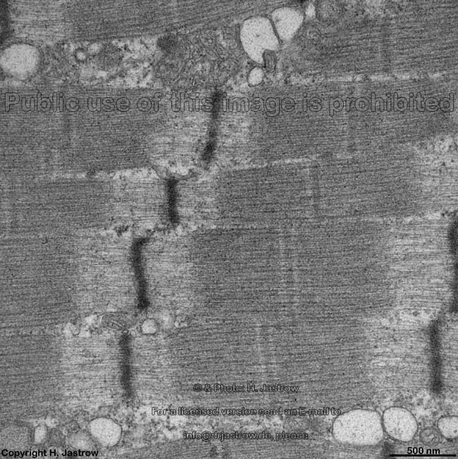
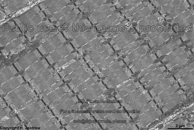
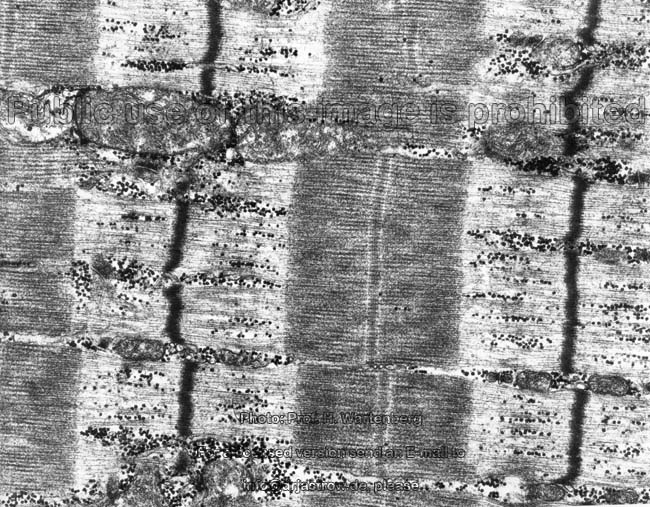
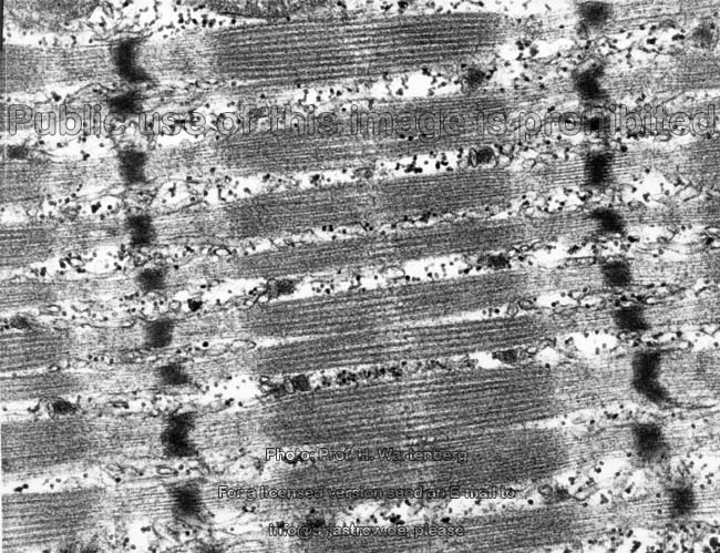
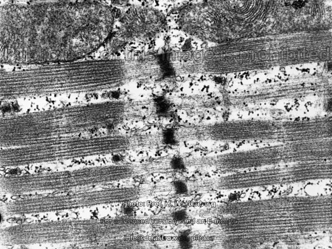
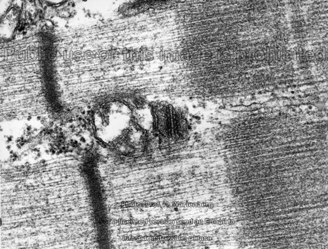
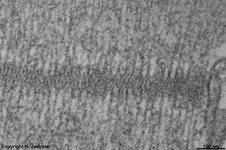
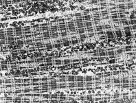
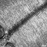
Z-linie
(rat) |
|
|
|
|
|
|
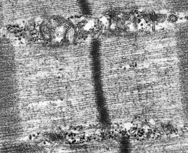
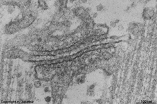
M-linie
(rat) |
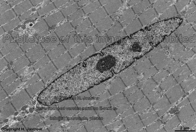
peripherical cell nucleus
(rat) |
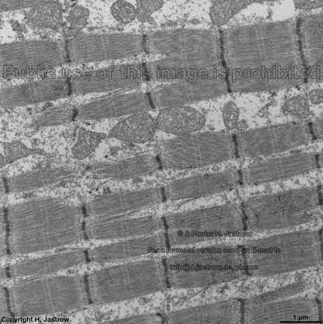
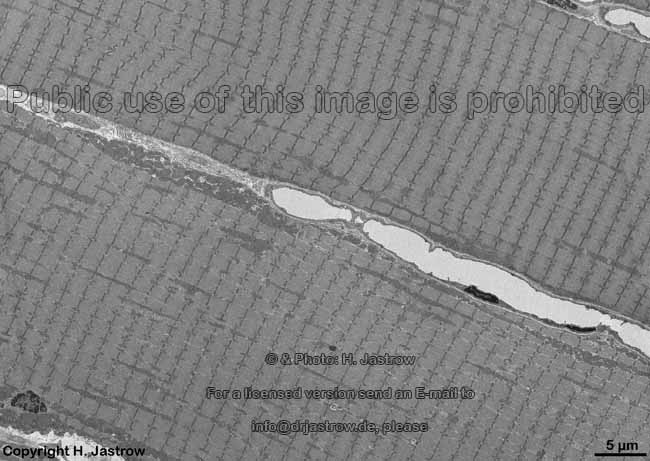
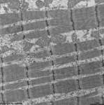
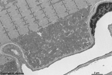
rump muscle fibres +
capillaries (rat) |
capillary with pores in
endothelium (rat) |
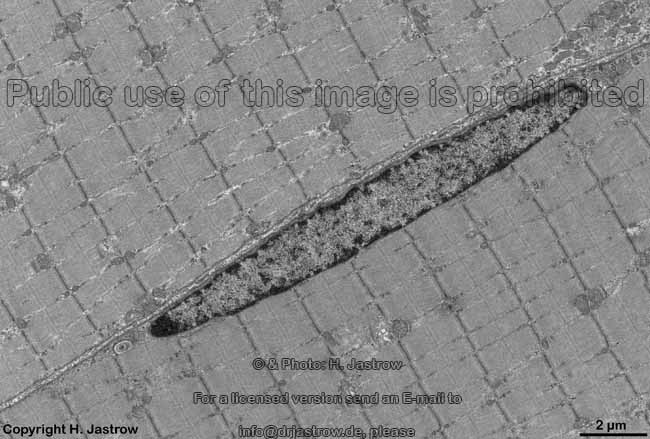
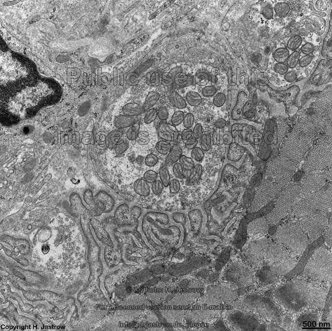
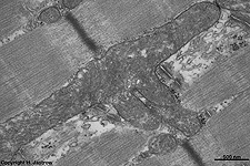
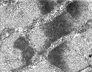
large aggregation of
mitochondria (rat) |
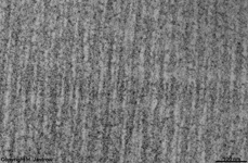
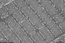
myofibrils
(rat) |
|
|
|
|
|
|
|
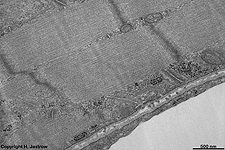
overview longitudinal
section 1 (monkey) |
longitudinal
section 2 (monkey) |
nucleus,
fibrils
(monkey) |
fibril bundles 1 (monkey) |
fibrils 2 (monkey) |
fibrils 3 (monkey) |
fibrils 4 (monkey) |
|
|
 |
|
|
|
|
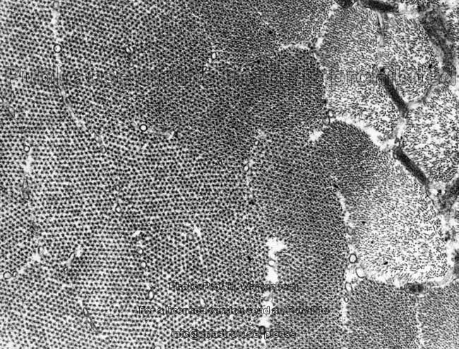
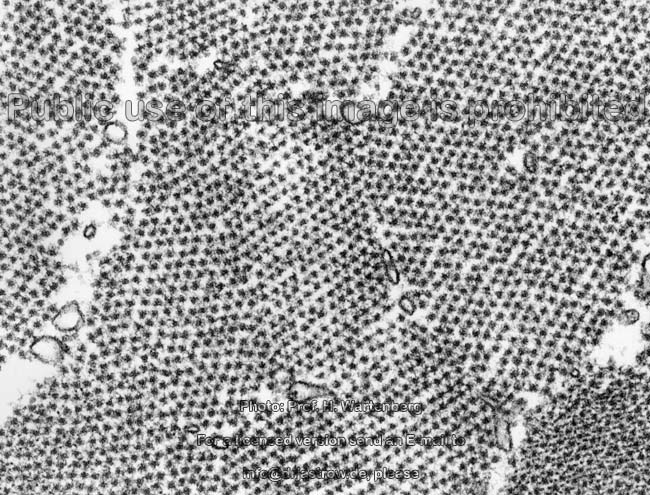
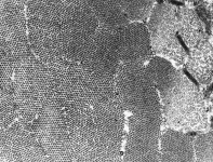
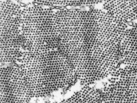
x-section of filaments
(monkey) |
cross-section: A-band,
M- and H-line (monkey) |
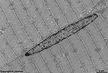
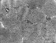
satellite cell, cross-section
(monkey) |
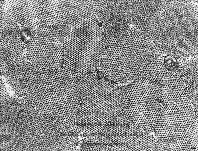
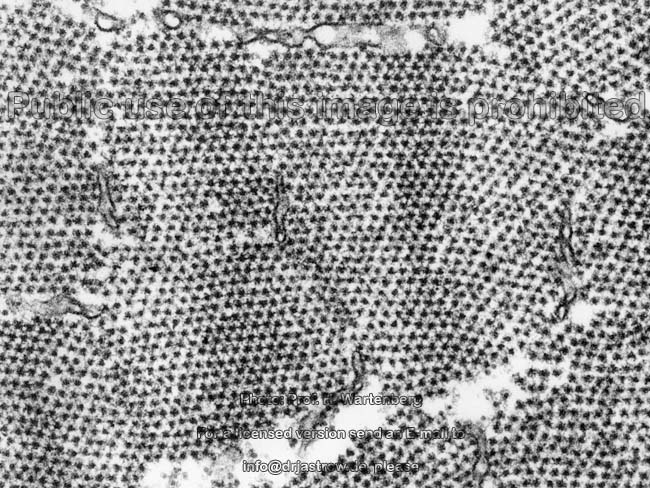
cross-section of Z-stripe
1 (monkey) |
cross-section of Z-stripe
2 (monkey) |
cross-section of Z-stripe
3 (monkey) |
cross-section of the
i-band (monkey) |
|
|
|
|
|
|
|
|
cross-section of A & i
band 1 (monkey) |
cross-section of the
M-stripe (monkey) |
cross-section of A & i
band 2 (monkey) |
cross-section of A & i
band 3 (monkey) |
sarkolemm,
T-tubule (monkey) |
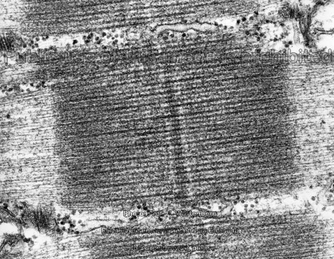
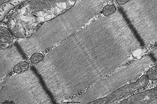
Z-stripe
(rat) |
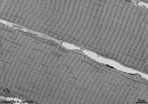
sarcomer (distance between
2 Z-stripes; rat) |
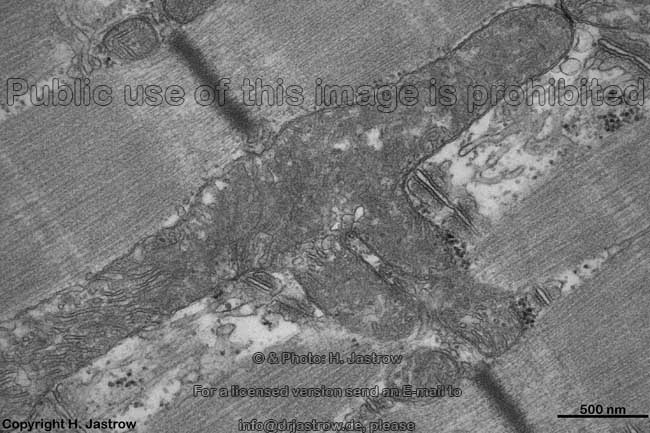
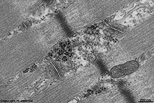
neuromuscular
synapse (rat) |
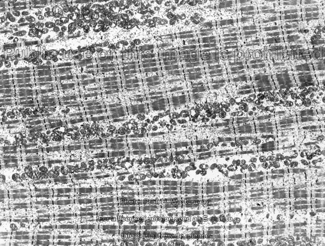
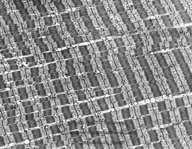
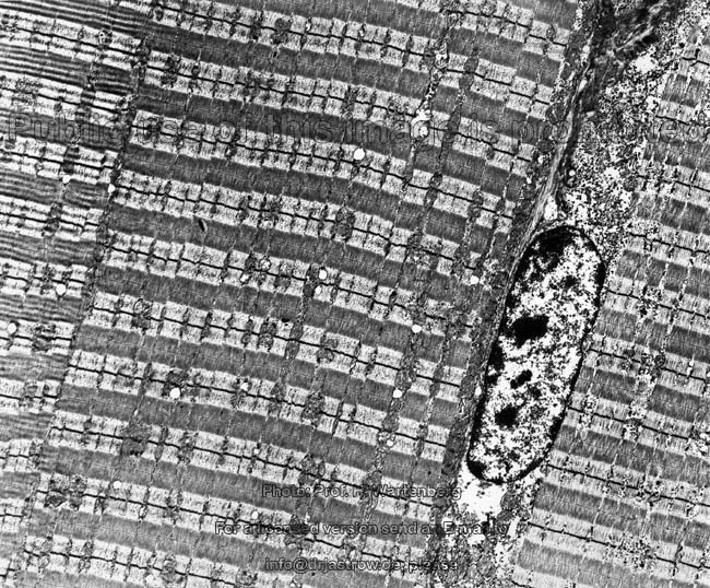
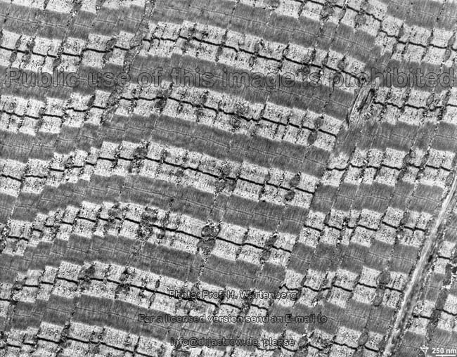
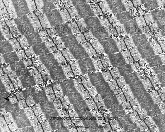
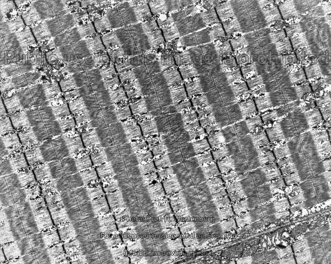
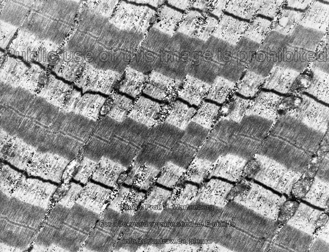
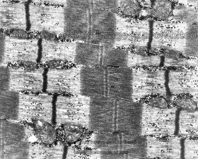
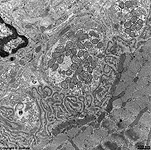
Skeletal striated muscle cells (Terminologia histologica:
Myocyti striatae non cardiaci) are extremely long (several centimetres)
and columnar in shape. Their diameters are between 10 and 100 µm,
in most cases 40 - 80 µm. This diameter increases under contraction.
Innumerable disc-like nuclei are always located to the margins of the cells
and lie close to the cell membrane which is called
here sarcolemma (Terminologia histologica: Sarcolemma). The
cytoplasm
of these muscle cells is called
sarcoplasm (Terminologia histologica:
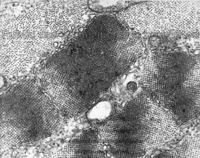
Sarcoplasma) contains some hundreds to thousands of myofibrils
(Terminologia histologica: Myofibrillae). The latter are round, several
centimetres long and have diameters of (0.1 to1.5 µm). They consist
of a periodic sequence of actin- and myosin
filaments in parallel order and associated proteins (see below).
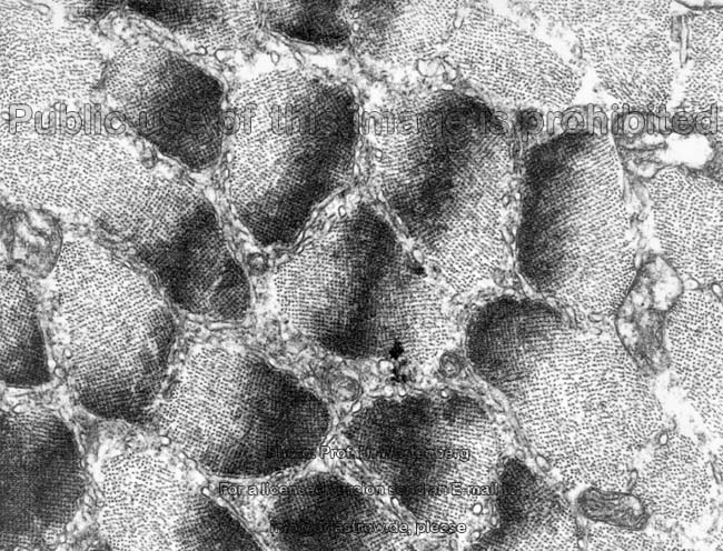
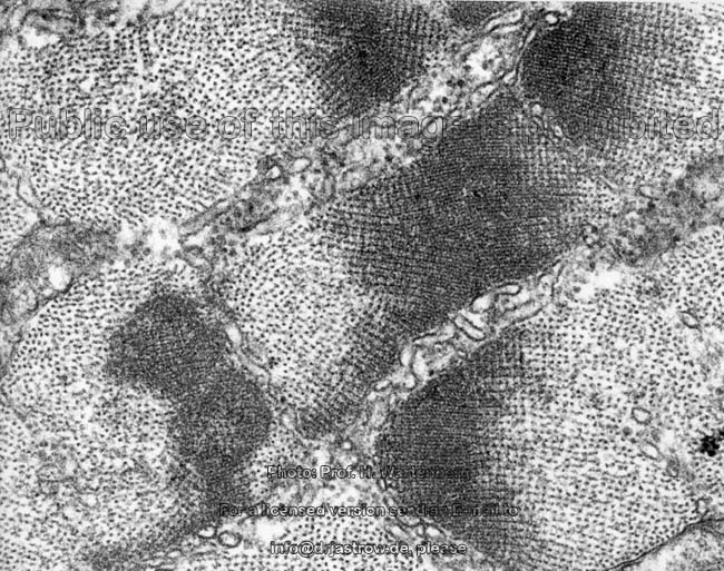
Usually very large aggregations of mitochondria
of crista-type lie in close vicinity. Further such mitochondria are seen
in a regular order along the myofibrils adjacent to the Z-disc region with
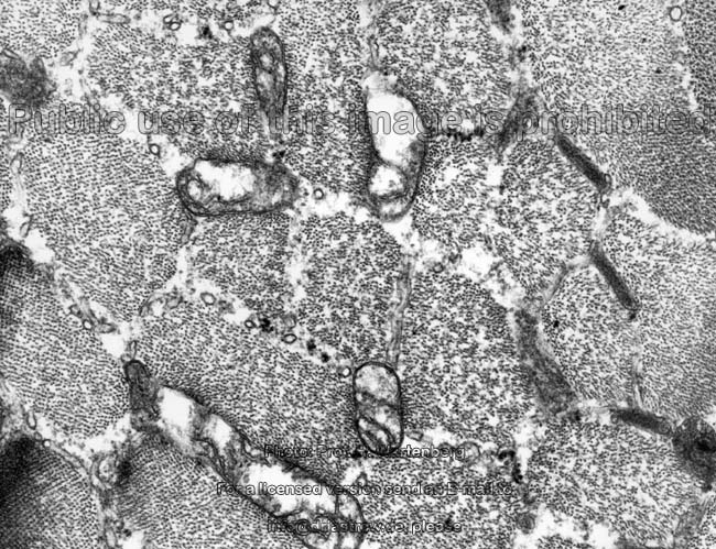
a central interruption caused by the very long T-tubules
(= transverse tubules; Terminologia histologica: Tubulus T; Tubulus transversus)
which extend as long tubular invaginations of the cell
membrane very deep into the cytoplasm.
These T-tubules are flanked on two opposing sides by long cisterns of the
smooth
endoplasmic reticulum (SER), which is called sarcoplasmic reticulum
(Terminologia histologica: Reticulum sarcoplasmicum) in skeletal muscle
cells. These cisterns of the SER are also termed longitudinal- or shorterL-Tubules
(Terminologia histologica: Tubulus longitudinalis, Tubulus L). These 3
adjacent tubules (L-T-L) which are termed Triads (Terminologia histologica:
Trias) are characteristic for skeletal musculature. The SER extends like
a net from the cisterns next to the T-tubules around the fibrils. It is
the essential storage site for calcium ions which are needed for muscular
contraction. Larger or smaller amounts (depending on the type of muscle
fibre) of beta-glycogen granules
are present next to the myofibrils as well. A considerable quantity of
invaginations and infoldings of the cell
membrane is seen on the long ends of muscle cells. They serve for surface
enlargement. The last actin filaments of the
myofibrils are directly connected to integrin molecules which reach through
the cell membrane and bind tightly to
the basal lamina which is
further connected to collagen fibrils of
an adjacent tendon or a periosteum
or a perichondrium.
Directly adjacent to the skeletal muscle
cells small mononuclear myosatellite cells (Terminologia histologica:
Myosatellitocyti) are present which are able to undergo mitosis whereby
one new cell later will be incorporated into the muscle cell contributing
its nucleus and cytoplasm since mitotic processes do not take place inside
the large muscle cells themselves. However for growing and under training
conditions a higher protein synthesis is required in muscle cells for increase
in thickness thus more genetic information, i.e. new nuclei are necessary.
The satellite cells are resting myoblasts located under the basement membrane
coat of the muscle cell.
An English page with much more detailed information is available in
the professional version of this atlas.
--> differential diagnosis of muscle tissues,
heart
muscle, smooth muscles, neuromuscular
junction,
L-tubules, beta-glycogen,
actin
filaments
--> Electron microscopic atlas Overview
--> Homepage of the workshop
Many images were kindly provided by Prof. H. Wartenberg;
other images, page & copyright H. Jastrow.
I am grateful to Dr. G. Spatkowski for the specimen of her eye muscle.