Overview glycogen granules (Granula
glycogeni):
Pages with explanations are linked to the
text below the images if available! (Labelling is in German)
 |
 |
 |
 |
 |
 |
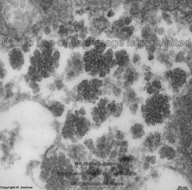
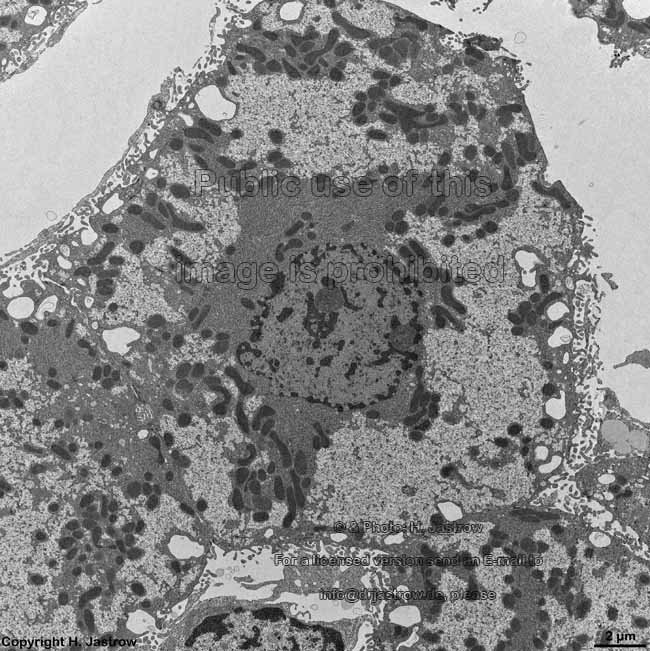
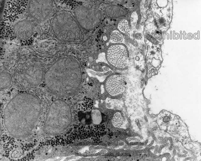
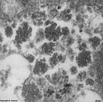
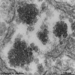
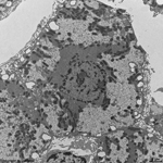
alpha-glycogengranules
of a liver cell 1 (rat) |
alpha-glycogengranules
of a liver cell 2 (rat) |
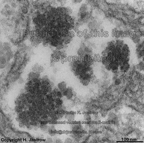
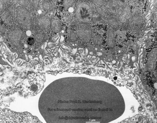
alpha-glycogen granules
of 2 liver cells (rat) |
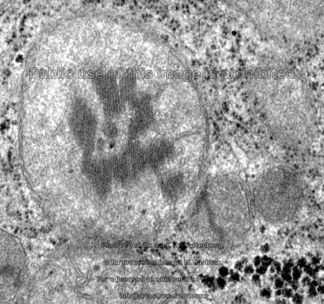
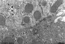
hardly stained* Glycogengra-
nules liver cell 1 (rat) |
hardly stained* Glycogengra-
nules liver cell 2 (rat) |
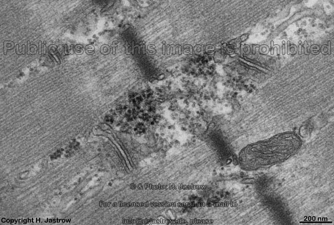
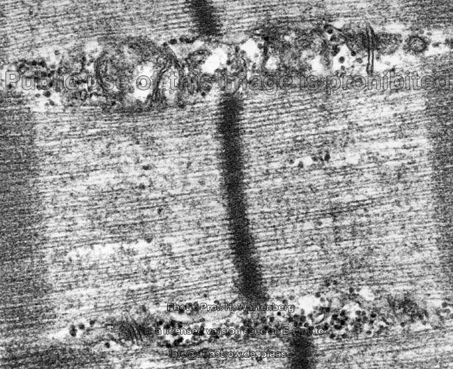
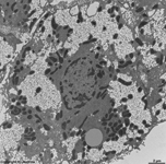
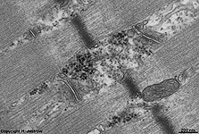
beta-Glykogen granules ske-
letal muscle cell 1 (rat) |
* modified contrast applied here to stain glycogen less intense. This allows
easy distinction from ribosomes.
Glycogen is a very large polymer only consisting
of glucose (homoglycane) mainly in alpha 1,4 glycosidic junction to
which enzyme complexes as well as cofactors for aggregation and
dissociation of further glucose units are attached. Helical chains
of alpha 1,4 glycosidicly connected glucose units bind further such
chains via 1,6 glycosidic junctions on every 8th to 12th glucose unit.
This is the molecular base of a branched three-dimensional network of glucose
units with about 12 levels of branching that finally forms the glycogen
particles. Further water is associated to the molecule, i.e. it is hydratised.
The spheroid shape of glycogen is optimized for space-saving compact storage
and quick possibility for enzymes to access the macromolecule for modification. The
attached enzymes areglycogenin, glucose-phosphate-mutase, UDP-glucose-phosphorylase, amylo-alpha-(1,4-->1,6)-transglycosylase, glycogen
synthase a and b, which serve for steps of glycogen synthesis
as well as glycogen phosphorylase, trisaccharid-transferase, alpha
1,6-glycosidase, glucose-phosphate-mutase
and glucose-6-phosphatase
which are involved in glycogenolysis.
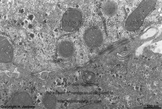
Glycogen storage granules (Terminologia histologica: Granula
glycogeni) are seen in the cytoplasm of
many cells. They always have irregular borders and are not limited by any
membranes. For light microscopic visualisation of glycogen either the carmin
staining according to Best (red staining
of cytoplasmic areas which contain glycogen) or PAS
staining is recommended which, however, also stains other sugars in red.
The following different types of glycogen granules can be differentiated:
1. an alpha-glycogen granule
(Terminologia histologica: Alpha granulum glycogeni) is a rosette-like
aggregation of 5 to over 40 single glycogen particles with resulting
diameters of 90 and up to over 200 nm, and
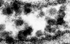
2. a much smaller single spherical beta-glycogen
granule (Terminologia histologica: Beta granulum) resembles
a single ribosome (diameter in sections about
15 - 20 nm) but is a little less electron-dense.
Diameters of beta-granules
are about 20 to 40, in most cases about 30 nm.
3. The rare gamma-glycogen granules
(Terminologia histologica: Gamma granula glycogeni) most probably are either
not fully synthesised or partly degraded glycogen granules with diameters
beyond
20 nm.
Whereas alpha-granules are most commonly seen in hepatocytes
where they are the morphological correlate for quick short-term storage
of large amounts of glucose during anabolic phases. The beta-granules are
typical for skeletal or heart
muscle cells which may also show few alpha-granules.
--> liver, ribosomes,
RER,
skeletal
muscle,
heart muscle
--> Electron microscopic atlas Overview
--> Homepage of the workshop
Five images were kindly provided by Prof. H. Wartenberg;
other images, page & copyright H. Jastrow.