Overview bones (Ossa)
and ossification (Ossificatio):
Pages with explanations are linked to the
text below the images if available!
 |
 |
 |
 |
 |
 |
 |
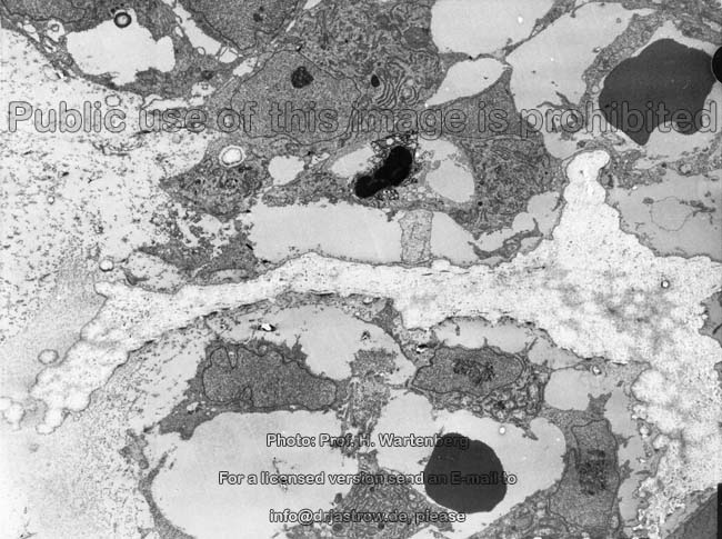
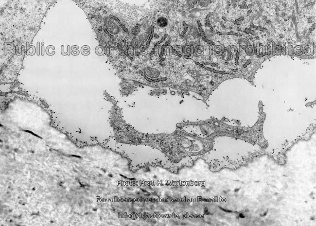
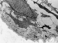
hardly active
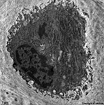
osteoblasts (monkey)
|
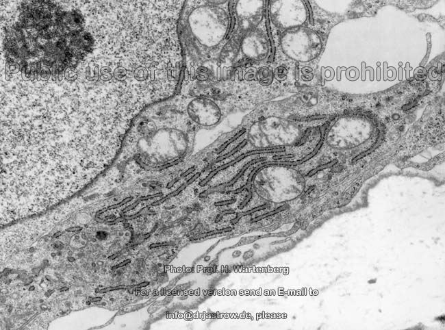
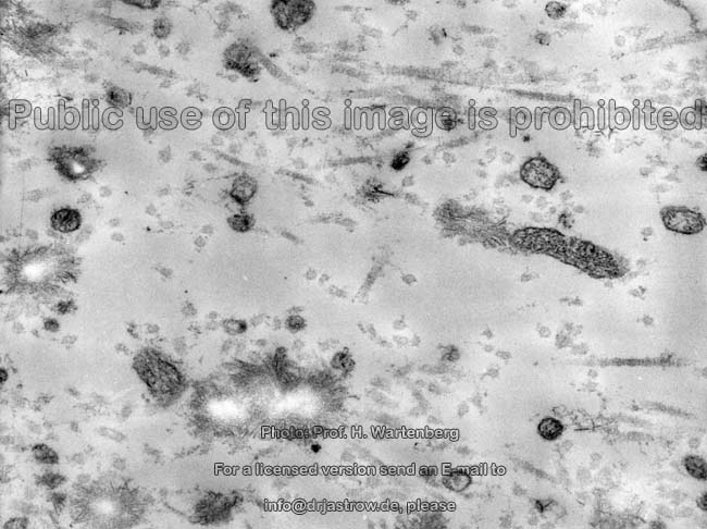
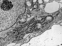
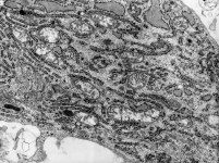
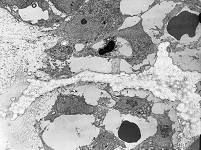
organells of an
osteoblast 1 (monkey)
|
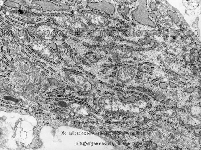
organells of an
osteoblast 2 (monkey)
|
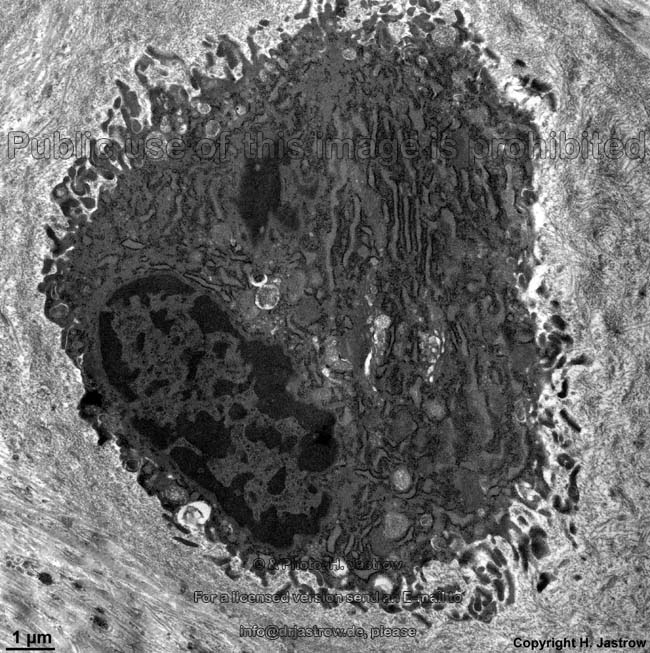
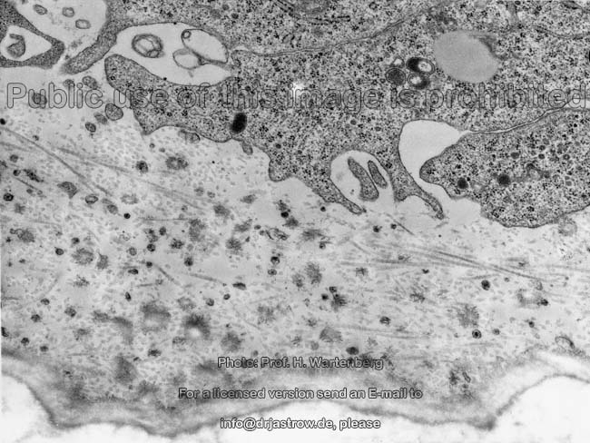
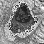
osteocyte
(rat) |
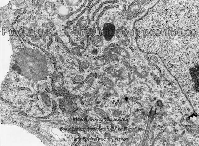
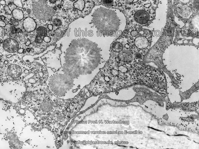
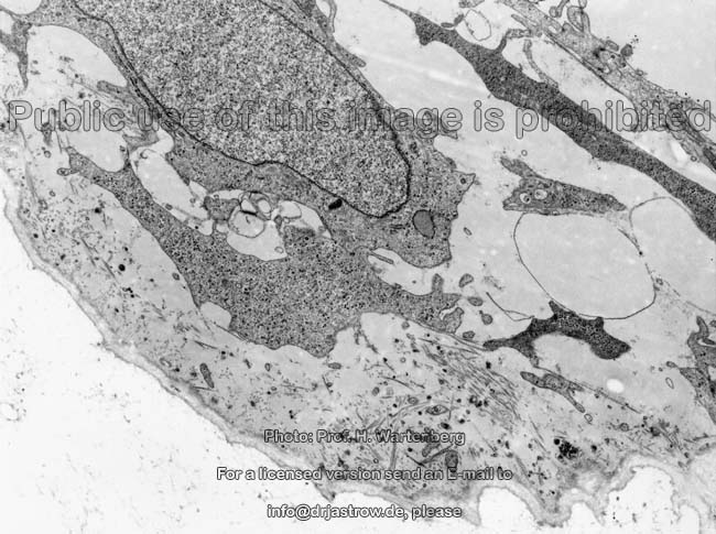
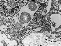
opening zone in chondral
offificattion(monkey)
|
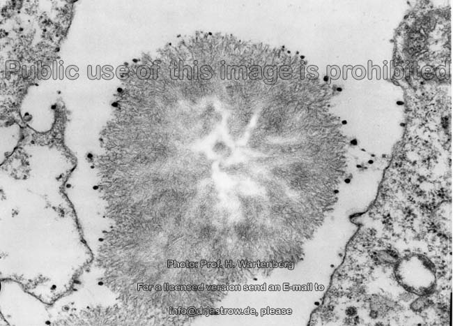
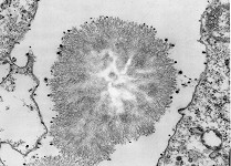
crystallisation centre
(monkey)
|
overview thereof |
 |
 |
 |
 |
 |
 |
|
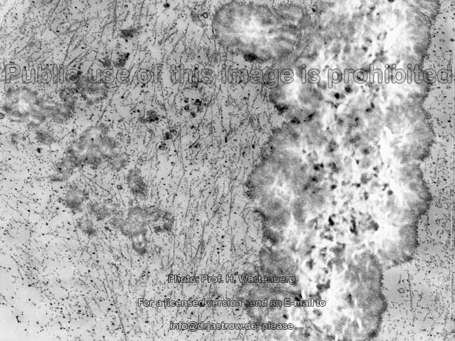
fresh osteoid (monkey)
|
osteoid formation 1
(monkey) |
fresh osteoid
detail (monkey) |
osteoid formation 2
(monkey) |
osteoid formation
3 (monkey) |
osteocyte
(rat) |
In humans the usually altogether 210 Bones (Terminologia
histologica: Ossa) consist of bone tissue
(Terminologia histologica: Textus osseus). Two main types of bones can
be distinguished: flat bones with
a massive, i.e. hard outer and inner layer and the elongate long
bones with large central cavities.
cells of bones:
all bone tissues consist of bone cells and the ossified matrix
around them. These bone cells are:
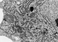
Osteoblasts (Terminologia histologica:
Osteoblasti) with a high metabolic activity. These cells build up bone
tissue as long as they are activated and not present as inactive lining
cells in the end- or periosteum.
Osteocytes (Terminologia histologica:
Osteocyti), the immured bone cells with low metabolic activity form dozens
of up to 200 µm long, 150 nm thin osteocyte processes (Terminologia
histologica: Processus osteocyti) and on the latter connexin
43-containing gap junctions establish connections
in between the cells and to endothelial cells
of Haversian canals. The
processes are located in tiny bone canaliculi (Terminologia histologica:
Canalculi ossis) with diameters up to 400 nm which interrupt the bone
matrix.
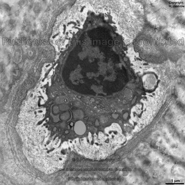
Osteoclasts (Terminologia histologica:
Osteoclasti) belong to the mononuclear phagocyte system despite the fact
that they always have many (up to 25) nuclei. They are giant cells
with diameters of 30 - 100 µm arising from fusing monocytes
from blood which are true mononuclear cells.
Numerous Golgi apparatuses produce numerous
vesicles
most of which are lysosomes. The latter release
their content by exocytosis in the
area of the ruffled border. Together with
the hydrochloric acid these substances destroy the bone
matrix.
Bone lining cells (Terminologia
histologica: Cellulae vestientes osseorum) mainly comprise inactive osteoblasts
resting in the
endosteum and periosteum
which are interconnected by gap-junctions forming
a flat epithelium since hardly
any intercellular substance
is present in between them.
Kinds of bone tissue:
1. Woven bone (Terminologia
histologica: Textus osseus reticulofibrosus) is the primary kind of
newly formed bone.
2. Trabecular bone (spongy
bone, cancellous bone; Terminologia histologica: Textus osseus spongiosus,
Textus osseus trabecularis) appears as a spongy framework with many holes
consisting of bone trabecula and depending on long-time pressure forces
is constantly remodelled.
3. Bundle bone (Terminologia
histologica: Textus osseus fasciculatus) forms the transition area from
trabecular
bone to lamellar bone showing larger bundles
of still lamellated bone which then terminate into the trabecula trabecular
bone nearly devoid of Haversian
systems.
4. Compact bone (Terminologia
histologica: Textus osseus compactus) lies as massive compact bone tissue
on and directly under the surface of long bones as well as Substantia
or Pars compacta on the bone shaft (diaphysis), or
as
Substantia corticalis on the ends of bones. Further, the inner
and the outer surface of skull bones (Tabula interna and - externa) consist
of stable Substantia corticalis. In the temporal bone (Os temporale) compact
bone tissue protects the structures of the inner
ear. Only in this area it is organised as woven
bone otherwise it is present in form of
Lamellar bone (Terminologia
histologica: Textus osseus lamellaris). The latter consists of 2-4 mm
strongbone lamellas (Terminologia histologica: Lamellae osseae). Hereby
in most cases lamellas of concurrent (aniostropic)
collagen
fibrils alternate with lamellas containing collagen
fibrils running in opposite direction which can be observed in a polarisation
microscope as texture with light and dark bands. The lamellas are usually
oriented longitudinally to the bone shaft.
Much more information and many more images are available in the professional
version of this atlas.
--> hyaline-, elastic
cartilage, connective tissue, resident
connective tissue cells, bone marrow
--> Electron microscopic atlas Overview
--> Homepage of the workshop
Most images were kindly provided by Prof. H. Wartenberg;
other images, page & copyright H. Jastrow.