Hardly active bone producing cells (osteoblasts) of a monkey
(for unlabelled original image click here,
please)
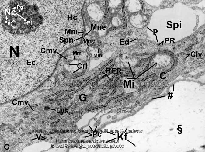
§ = mineralised bone (extracellular
matrix with calciumhydroxylapatit crystals);
# = border seam (in this small region
tiny crystals are being integrated into the non-calcified matrix called
osteoid);
C = cytoplasm (cellular
fluid with organells); Clv = Vesiculum clathrinum (clathrin-coated
endocytotic vesicle);
Cmv = Corpusculum multivesiculare (multivesicular
body);
Cri = Crista mitochondrialis (infolded inner membrane of a mitochondrium);
Ec = euchromatin;
Ed = Endocytosis (intake
of substances by formation of a Clv); G =Complexus golgiensis
(Golgi-apparatus);
Hc = Heterochromatin;
Kf
= Fibrillae collagenosae (collagen fibrils;
mainly of type 1);
Lys = Lysosoma (heterolysosome);
Mi = mitochondria (crista-type);
Mm = Membrana mitochondrialis (mitochondrial
membrane); Mme = Membrana mitochondrialis externa (outer mitochondrial
membrane);
Mmi = Membrana mitochondrialis interna (inner mitochondrial
membrane, which infolds forming Cri);
Mne = Membrana nuclearis externa (outer nuclear
membrane); Mni = Membrana nuclearis interna (inner nuclear
membrane);
N = Nucleus (nucleus); Nc
= Nucleolus (nuclear body with 3
different regions: Pa, Pf and Pg);
P = Plasmalemma (cell membrane);
Pa = Pars amorpha nucleoli (amorphous part of the nucleolus);
Pc = Processus cellulares (immotile cellular processes); Pf
= Pars fibrosa nucleoli (fibrillar part of the nucleolus);
Pg = Pars granulosa nucleoli (granular part of the nucleolus);
PR = Polyribosomae (polyribosomes = grouped free ribosomes);
RER = rough endoplasmic reticulum;
Spi = Spatium intercellulare (intercellular
space mainly containing water); Spn = Spatium perinucleare (perinuclear
space);
Vs = Vesicula secreti (secretory
vesicle containing fine granular material which will contribute to
the extracellular matrix).
In contrast to other images of bone
formation (ossification) virtually no organic extracellular material
(osteoid) is located in front of the border seam in the present image.
This and the only small amount of RER indicates a very moderate
activity of the shown osteoblasts. The mineralised bone (§)
has nearly no electron-density and shows only very few shadows of
collagen fibrils (Kf).
Nevertheless, it consists of a dense network of such fibrils to which calciumhydroxylapatit
crystals are attached as main component. This connection process takes
place in the region of the border seam (#).
--> bone, connective
tissue
--> Electron microscopic atlas Overview
--> Homepage of the workshop
The image was kindly provided by Prof. H. Wartenberg,
page & copyright H.
Jastrow.


