Overview heterochromatine (Heterochromatin):
Pages with explanations are linked to the
text below the images if available! (Labelling is in German)
An entire English version of this page is
in preparation!
 |
 |
 |
 |
 |
 |
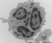
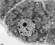
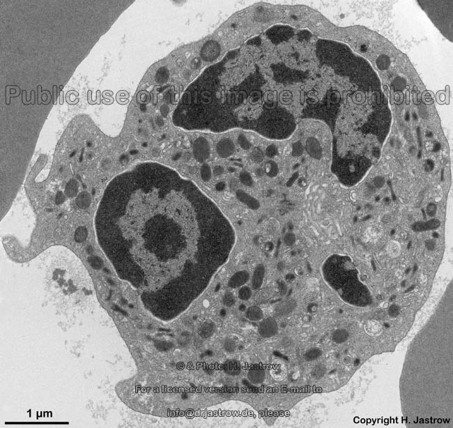
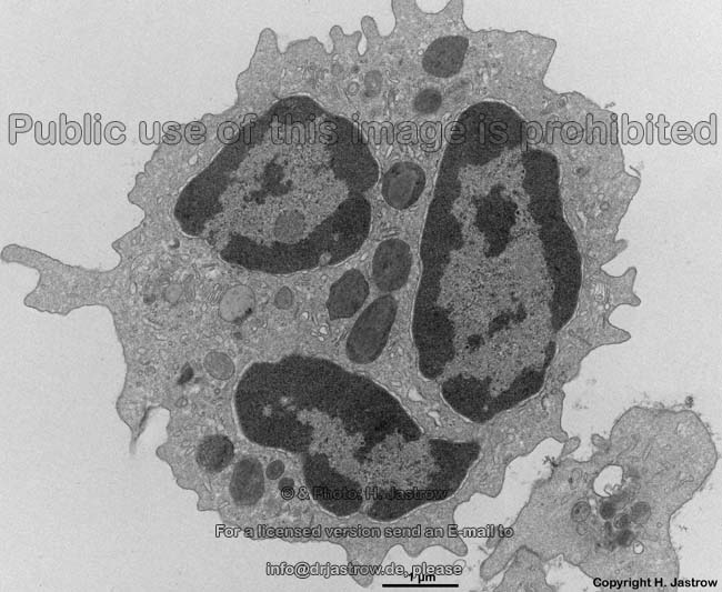
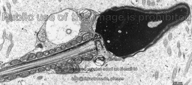
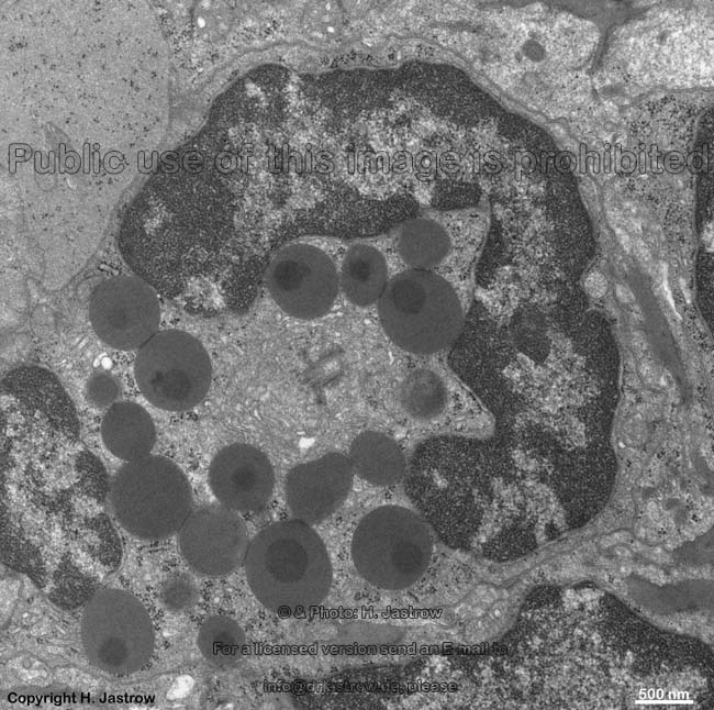
segmented nucleus of
a human eosinophil |
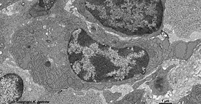
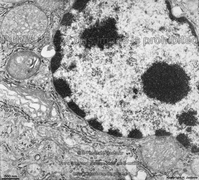
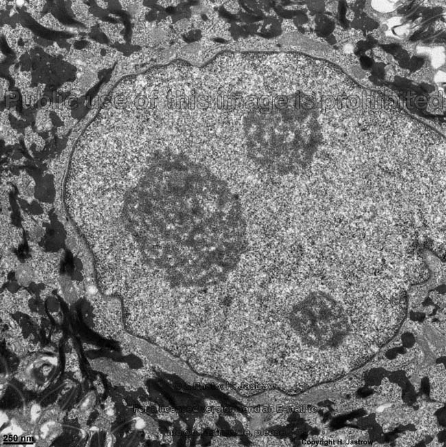
typical orientation of euchromatin as
"spoke of a wheel" in a plasma cell (rat) |
"spoke of a wheel" oriented
euchromatin in a human plasma cell |
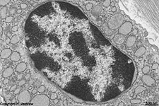
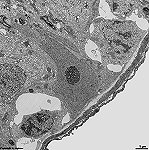
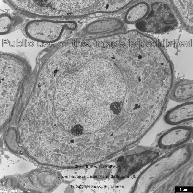
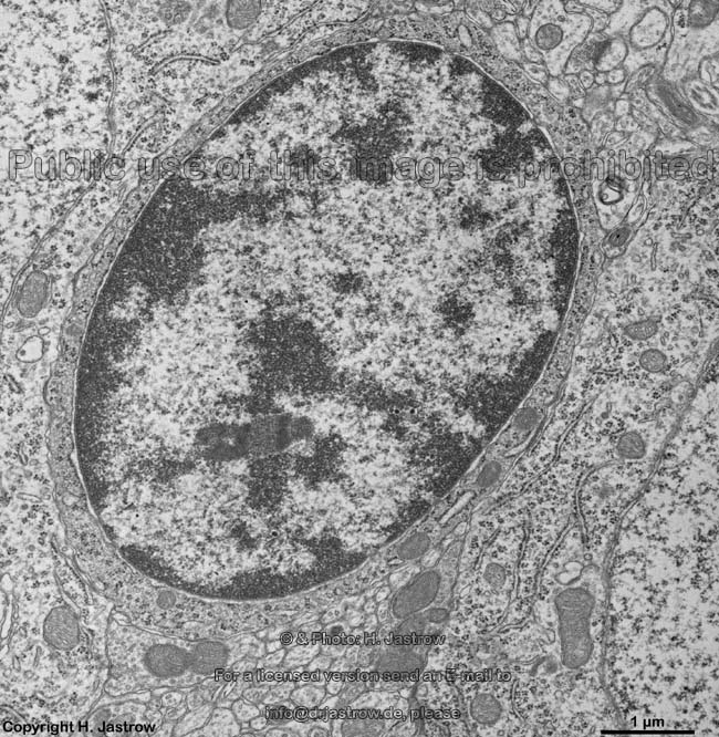
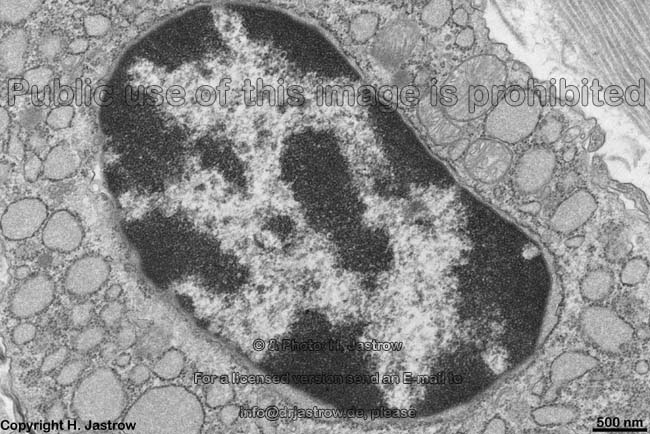
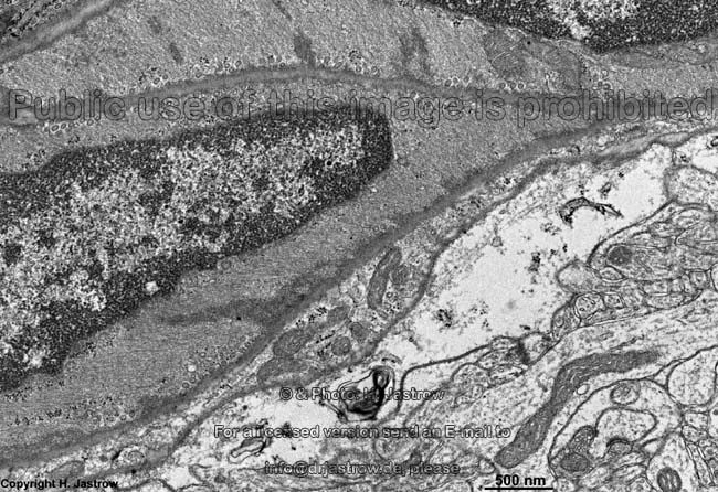
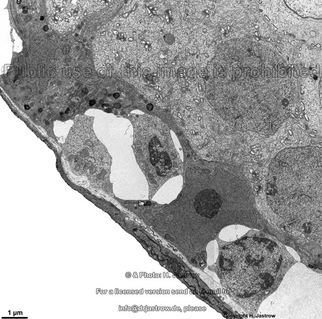
nucleus of a smooth muscle
cell (rat) |
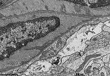
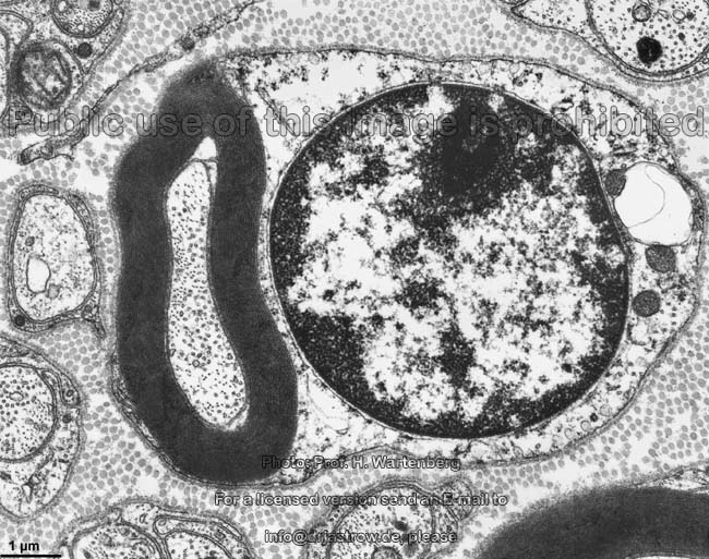
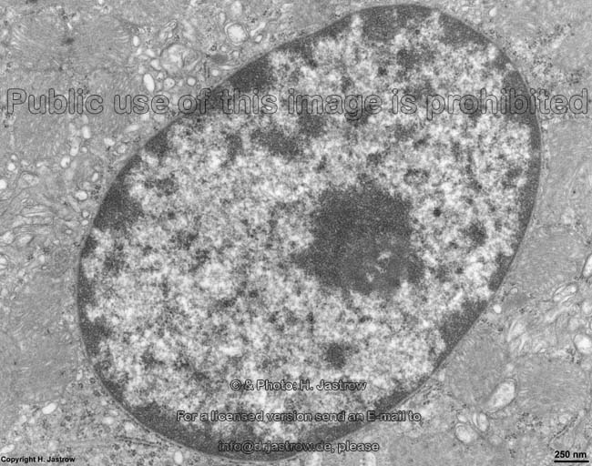
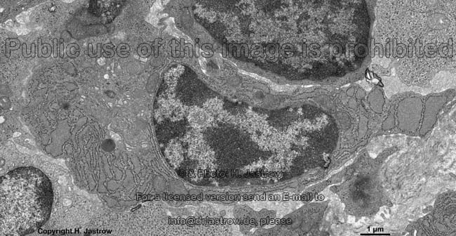
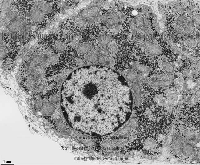
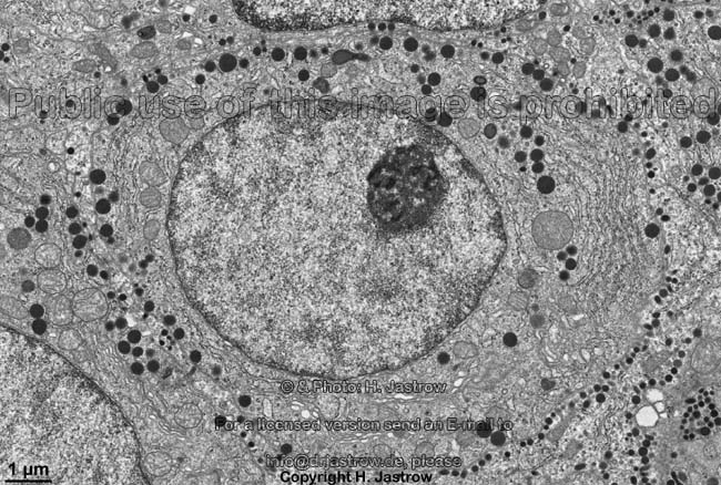
hepatocyte nucleus
(monkey) |
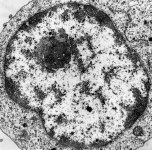
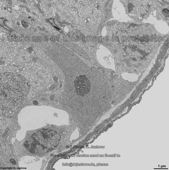
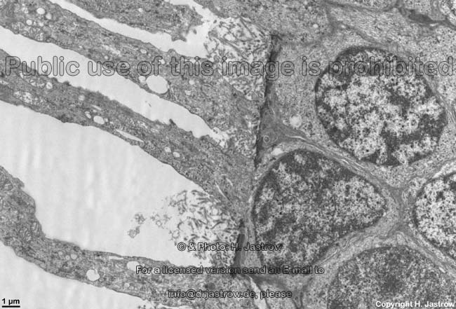
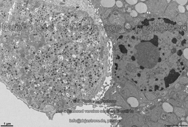
nucleus of a Sertoli
cell, testis (rat) |
The heterochromatin (Terminologia histologica: Heterochromatinum)
comprises the dense areas of chromatin (Terminologia histologica:
Chromatinum) in the karyoplasm of a cell.
Due to its high absorption of electrons it apperas dark, i.e. electron-dense
in transmission electron microscopic images. Heterochromatin is basophilic
in light microscopy. In heterochromatin desoxyribonucleic acid (DNA) is
present in spiral form, i.e. it is bound to histon-
and non-histon piation will always remain unused and are termed constitutive
heterochromatin (missing in Terminologia histologica; proposal: Heterochromatinum
constitutivum). In contrast,
the facultative heterochromatin (missing in Terminologiroteins
(see chromosomes). Thus the DNA is present
here only as double-helix and cannot be read, i.e. it is inactive genetic
information. Those sections
of chromosomes which in a cell will never
be transcribed due to its differenta histologica; proposal: Heterochromatinum
facultativum) comprises chromosomal regions
which are only transcribed during short, limited time intervals. Cells
with high content of heterochromatin and only few euchromatin
have a low synthetic activity (e,g., fibrocytes)
or only a mall portion of the genetic information is necessary to be transcribed
for proper cell function as, e.g., in case of plasma
cells which secrete only one type of immune
globuline. Condensations
of chromatin, i.e., heterochromatin often is associated to the nucleolus
(nucleolus-associated heterochromatin; missing in Terminologia histologica;
proposal: Heterochromatinum adjunctum nucleoli) or
it is attached to the nuclear
lamina (nuclear lamina associated heterochromatin; missing in
Terminologia histologica; proposal: Heterochromatinum adjunctum laminae
nuclearis). There is a very
characteristic distribution of eu- and
heterochromatin in plasma cells that reliably
allows cell identification: the spokes-of-a-wheel chromatin structure,
wherby the spokes are formed by the lighter euchromatin
which then in ideal case runs from a central dark nucleolus
towards the nuclear pores. Small
areas of very electron-dense heterochromatin which mainly are surrounded
by euchromatin are termed chromatin
granules (Terminologia histologica: Granumum chromatini).
Only in females about 25 - 30% of all nuclei
show a very considerable lump of heterochromatin associated to the nuclear
lamina in light microscopy, the sexchromatin (Barr's body;
Terminologia histologica: Chromatinum sexuale). The latter comprises an
inactive
and thus condensed X-chromosome
which is connected to the nuclear
lamina with both of its terminals (telomer regions). From the beginning
of somite formation on day 20 of embryonic development one and the same
X-chromosome
of each cell and daughter cells gets inactivated. In about 3% of all neutrophilic
granulocytes of females this condened X-chromosome
appears as drop- or drum-like appendix, i.e. drumstick on one of the nuclear
segments (it is also called Chromatinum sexuale in the Terminologia histologica). Only
with help of Quinacrin fluorescence staining it is possible to visualise
a small spot called Y-chromatin
(missing in Terminologia histologica; proposal Chromatinum Y) on the nuclear
membrane in males which corresponds to a large heterochromatine part of
the Y-chromosome.
--> nucleus, nuclear
membrane,
nuclear pore,
nucleoplasm,
euchromatin,
nucleolus,
chromosomes
--> Electron microscopic atlas Overview
--> Homepage of the workshop
Three images were kindly provided by Prof. H. Wartenberg;
other images, page & copyright H. Jastrow.


