Overview nucleolus (Nucleolus):
Pages with explanations are linked to the
text below the images if available! (Labelling is in German)
 |
 |
 |
|
 |
 |
 |
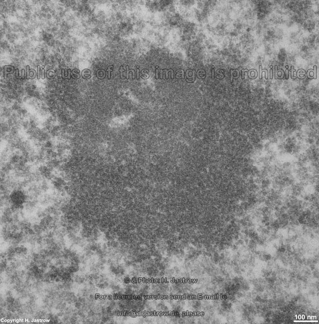
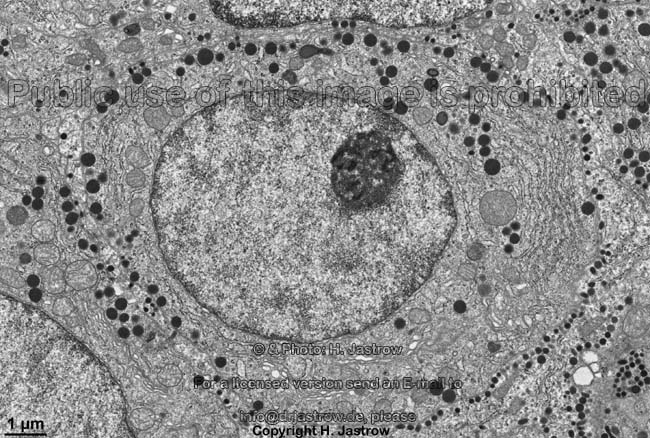
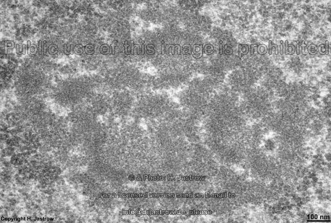
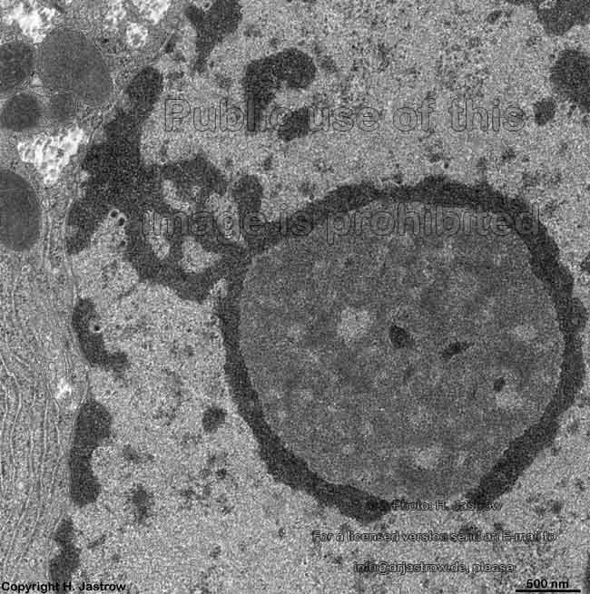
nucleolus of a neuron in
brain cortex (rat) |
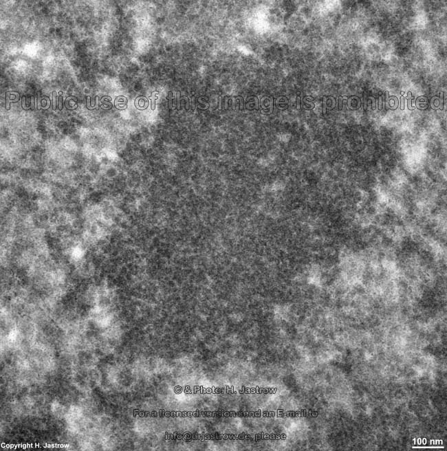
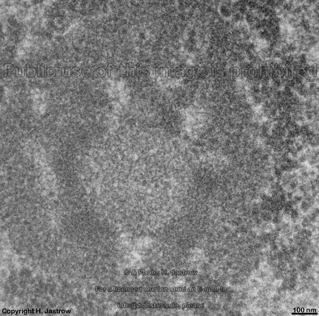
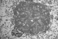
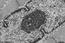
nucleolus of a mucous
salivary gland cell (rat) |
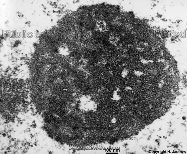
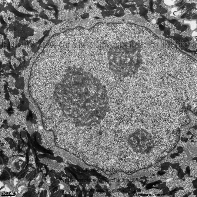
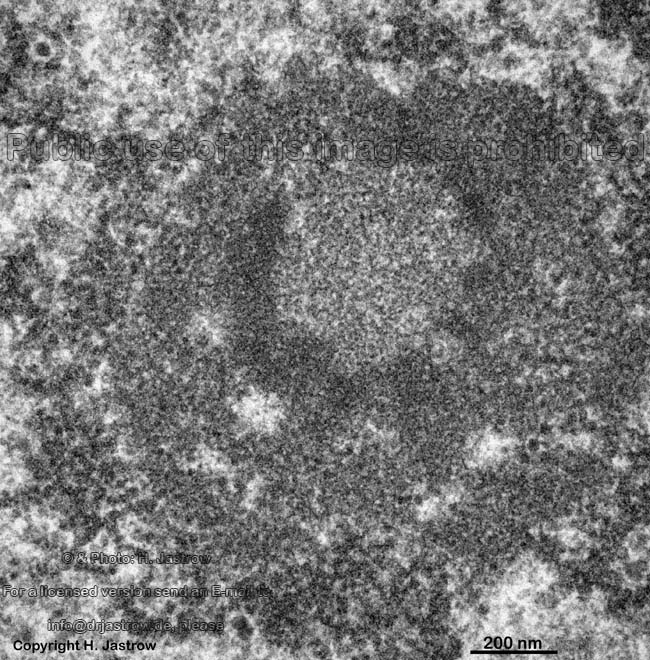
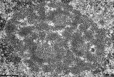
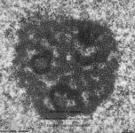
nucleolus of a
human plasma cell |
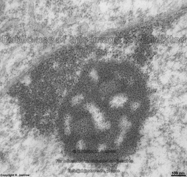
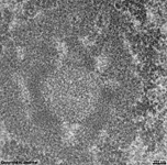
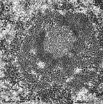
nucleolus Tonsilla
palatina (human) |
nucleolus skeletal mus-
cle cell nucleus (pig) |
nucleolus
Hypophysis (rat) |
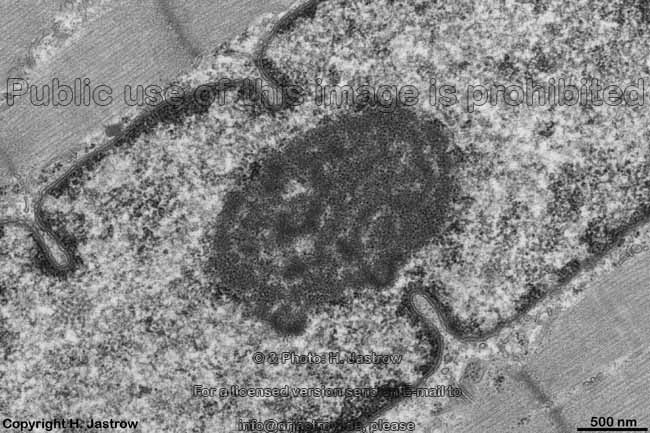
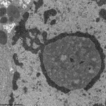
nucleolus of a
hepatocyte (rat) |
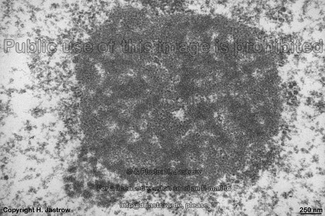
Nucleols (Terminologia histologica: Nucleoli) are spherical
to ovoid electron-dense basophilic condensations in the nucleus
of a cell with diameters of 1 - 2 µm. Nucleols are
the construction sites of ribosomes from
ribosomal subunits and may take up to ~25% of the nuclear
volume. Nucleols often are located in the centre of cell nuclei,
however, they also may be associated to the nuclear
membrane. They appear
next to nucleolar organizing regions (Terminologia histologica:
Nucleolum operans regiones) on secondary stranglings of the short arms
of the acrocentric chromosomes
13, 14, 15, 21 and 22. Since the latter are present twice in each cell,
theoretically a maximum of 10 nucleols is possible in human
cells, however in practice this is never the case. This is due to the fact
that the synthesis of sufficient ribosomal ribonucleic acid (r-RNA) even
in case of high demand is only possible at 2 to 3 nucleols, since several
nucleolar organizing regions then aggregate in large common nucleols. Plenty
of copies of the genetic information for formation of 5-S, 5,8-S, 18-S
and 28-S subunits of ribosomes are present
in such argyrophilic, i.e. stainable with sliver salts, nucleolar organizing
regions of the previously mentioned chromosomes.
Resting cells may show only small or even
no visible nucleols. Nerve cells, which
no longer undergo mitosis, in many cases only
show one very large nucleolus comprising all nucleolar organizing regions
of the whole nucleus. The shorter the interphase
during mitosis the higher is the number of
nucleols in quickly dividing cells. Since immediately after mitosis
all 10 nucleolar organizing regions form smaller nucleoli which then accumulate
to few larger ones. In many cases nucleols are connected to the inner
nuclear membrane via electron-dense chromatin bridges or are directly
attached to the nuclear membrane.
nucleols show several
1. fibrillar centres (Terminologia
histologica: Centra fibrillares) of low to slight electron density,
2. electron-dense fibrillar components
(Terminologia histologica: Partes fibrillares densae) with a delicate inner
structure,
3. a granular component (Terminologia
histologica: Pars granulosa) of the nucleolus. Additionally the nucleolus
contains
4. some small non-electron-dense regions
(amorphous part of nucleolus, nucleolar interstice; Terminologia
histologica: Pars amorpha nucleoli).
An English page with much more detailed information is only available
in the professional version of this atlas.
--> ribosomes, nuclear
membrane, nuclear pore,
karyoplasm,
euchromatin,
heterochromatin,
chromosomes,
nucleus
--> Electron microscopic atlas Overview
--> Homepage of the workshop
Images, page & copyright H. Jastrow.