Overview sebaceous glands (Glandulae
sebaceae):
Pages with explanations are linked to the
text below the images if available! (Labelling is in German)
The majority of sebaceous holocrine glands (Terminologia
histologica: Glandulae sebaceae holocrinae) are located in the areated
skin of the whole body. Here they
are usually associated to hair follicles delivering their secretion,
i.e. sebum into the dermal papillary
canal. Thus they are called hair sebaceous glands (Terminologia
histologica: Glandulae sebaceae adnexa pili). An example of such a gland
is the gland of Zeis (ciliary
sebaceous gland; Terminologia histologica: Glandula sebacea ciliaris) a
small sebaceous gland associated to the follicles of the eyelashes. Further,
there are free sebaceous glands (Terminologia histologica: Glandulae
sebaceae liberae) appearing with no relation to hairs. The
latter are encountered on the following locations: in the outer acoustic
meatus of the ear, in the outer region of the
lips (where the latter in contrast to the red margins are not mechanically
altered), in the area of the mamilla,
on the glans of the penis, in the skin
covering the labia minora and
around the anus. The largest of these free glands is the gland of Meibom,
a branched alveolar sebaceous
gland with wide end pieces connected
to a central excretory duct.
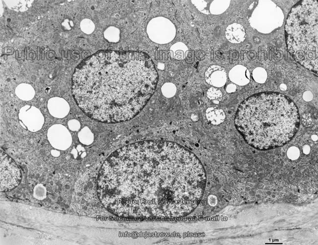
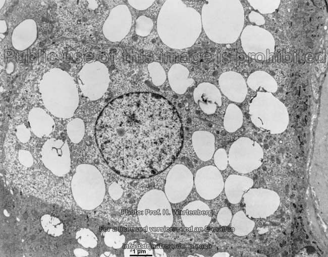
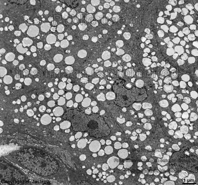
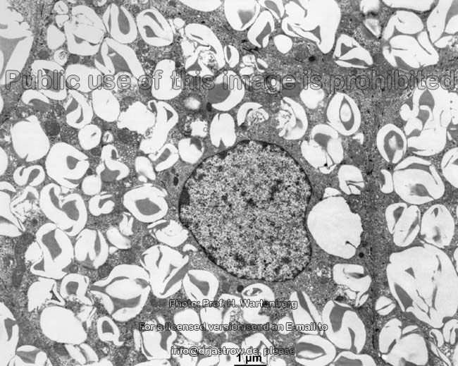
Sebaceous glands consist of differentiated epithelial
cells (sebaceous gland cells; sebaceous epithelial cells; Terminologia
histologica: Exocrinocyti sebacei; Epitheliocyti sebacei; Sebocyti). Their
wide end pieces called alveols
contain a stratified non-keratinised
epithelium. Peripheral basal cells
(Terminologia histologica: Cellulae basales periphericae) are located on
the basement membrane of these glands
and here constantly undergo mitosis whereby
one daughter cell remains on place while the other is pushed into the lumen.
Due to further proliferation of underlying cells it slowly reaches the excretory
duct (Terminologia histologica: Ductus excretorius). Hereby the cells
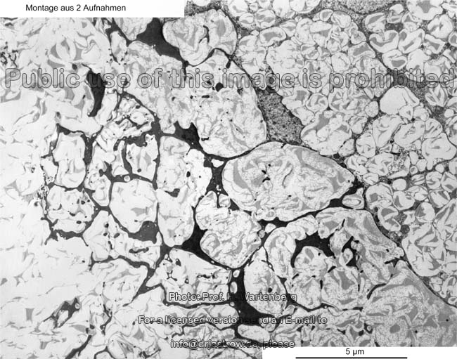
show more and more lipid droplets
that are confluent when becoming aggregations of sebum a special
composition of lipids. The nuclei as well as
the organelles of the maturing cells slowly
degenerate. Therefore the cells now are called vacuolated degenerating
cells (Terminologia histologica: Cellulae vacuolatae degenerantes. Thus
sebaceous glands secrete degraded cells proper, which comprise the
sebum, through their ducts. In case of the hair associated sebaceous glands
the secretion is pushed into the dermal
papillary canal and slips to the surface next to the hair.
Sebaceous glands produce the sebum
which makes the skin and hairs
soft and shiny. It is water rejecting and inhibits proliferation of bacteria.
The main components of sebum are different triglycerids and squalen.
The coryne bacteria of the skin degrade the sebum to fatty acids which
are partly responsible for the acid milieu on the surface of the
skin.
Sebaceous glands are simple
of branched alveolar glands,
i.e. the lumina of their glandular
saccules (Terminologia histologica: Sacculi glandulares) are wide.
The diameter of hair associated sebaceous glands is about 1mm.
--> glands, skin,
hair,
epithelium,
lipid
droplets
--> Electron microscopic atlas Overview
--> Homepage of the workshop
Four images were kindly provided by Prof. H. Wartenberg;
other image, page & copyright H. Jastrow.