Fig.1
 |
Ladies and gentlemen,
in the present information society with high-speed
computers and increasing network connections throughout the whole world,
medical education and particularly anatomy cannot do without the new media.
In this context the internet seems to be the best place to offer easily
accessible, interactive teaching modules for acquisition of a three-dimensional
functional picture of the human body. |
Fig.2
 |
Fig.3
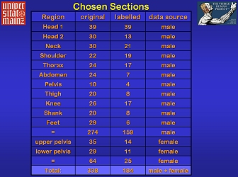 |
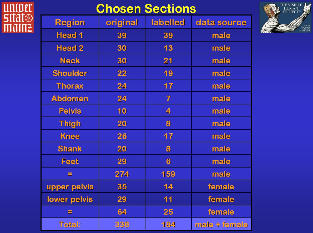
The atlas is based on digitised sections of the
Visible Human male and female as, at present, the most important international
source for free high quality images of human gross anatomy including CT
and MRI. In regions of special clinical interest like the head and with
many small details more sections were selected than in others with less
evident changes between the slices (Fig.3). Further, corresponding
axial radiological images were included.
Image data were obtained from the NLM
or its mirror
sites. Decompression and conversion into JPG files were necessary to
put the images at disposal in the internet. Thereby the original resolution
was maintained. 12-Bit greyscale CT- and MR images were reduced to 8-Bits
of grey. The JPG compression resulted in a strong reduction of the file
size without naked-eye detectable loss of colour contrast (Fig.4). |
Fig.4
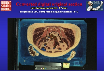 |
Fig.5 (link to labelled image atlas
page)
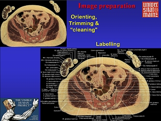 |
The converted pictures were oriented according
to radiological standard, trimmed and prepared to appear on a black background
(Fig.5). For labelling black margins were added in order not to
cover the pictures by the letters. Labelling was performed mostly by instructed
students attending the workshop according to the present international
terminology, Terminologia Anatomica, by using different anatomical atlases
and books. After thorough checks, revision, correction and trimming, internet
pages were composed.
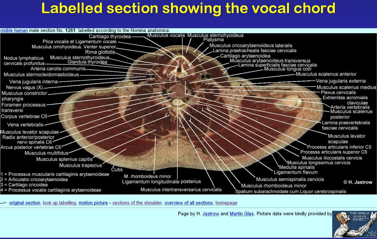
In Fig.6 you see such a web page. As many
structures as possible and reasonable were labelled on the pages with marked
sections. When a structure was not labelled, it was either not clearly
visible or there was not sufficient space available. In that case, the
structure was labelled on the neighbouring sections. There is no strict
uniformity in the layout, thus all labelled images reflect the individuality
of the involved student whose name is given below the image. |
Fig.6 (link to labelled image atlas page)
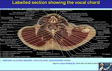 |
Fig.7 (link to atlas page)
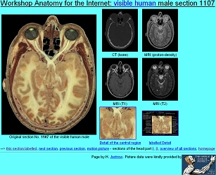 |
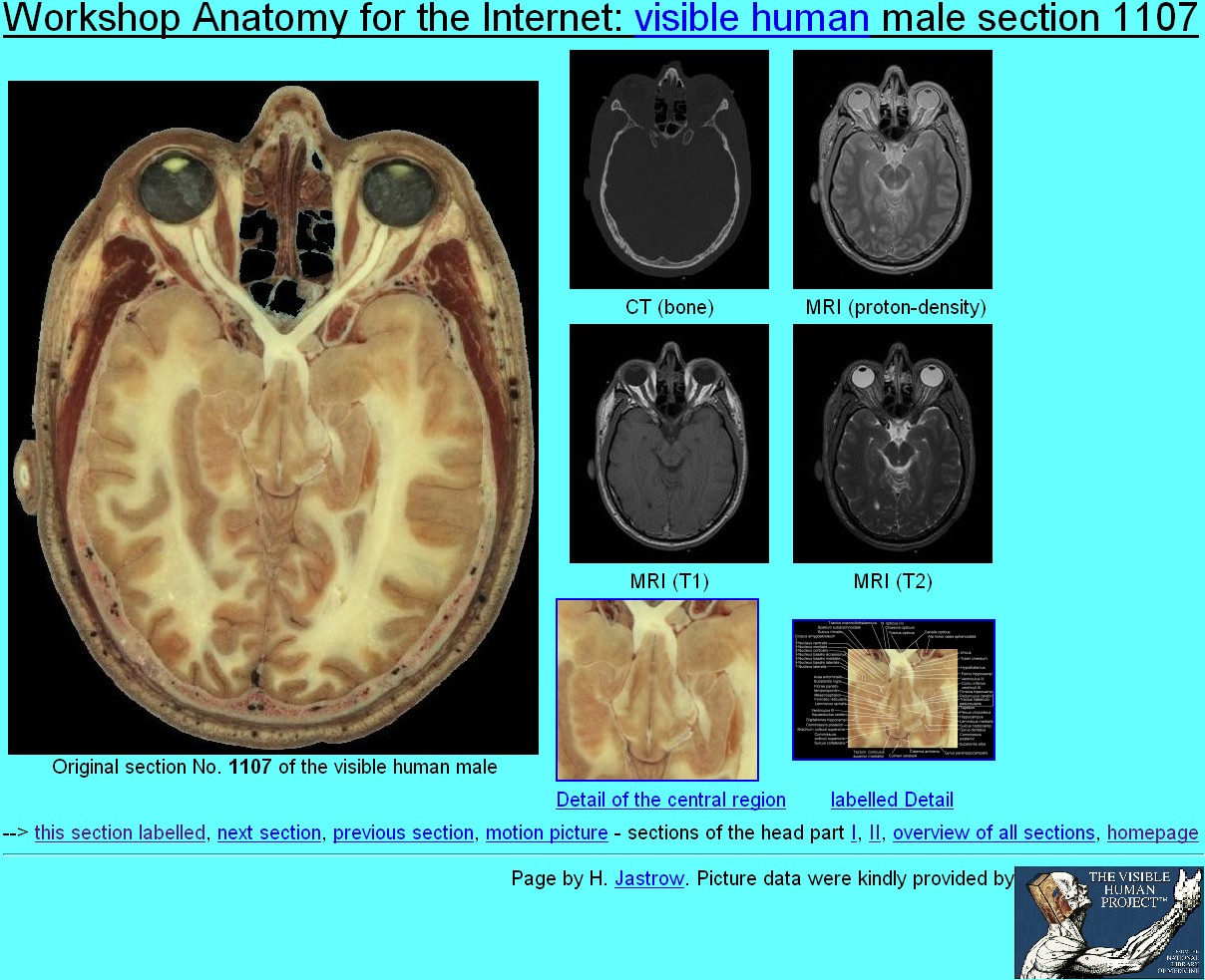
As you can see in Fig.7 images are presented
in full original pixel resolution in all pages of original sectionsin
the head, neck and lower limb regions. Prepared
sections, available corresponding CT and MR-images, and in some cases icons
of details enlarged on linked pages were arranged with further links using
the Netscape Composer.
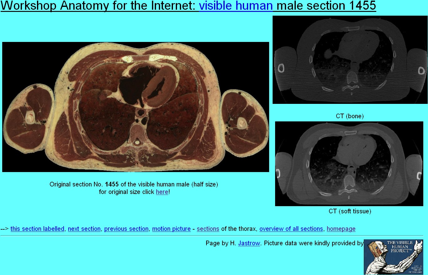
The images of pages with sections of the thoracic,
abdominal and pelvic regions were reduced to half size with a link to the
full-sized picture in order not to push the CT scans out of the screen
(Fig.8). |
Fig.8 (link to atlas page)
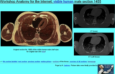 |
Fig.9 (link to atlas page)
 |
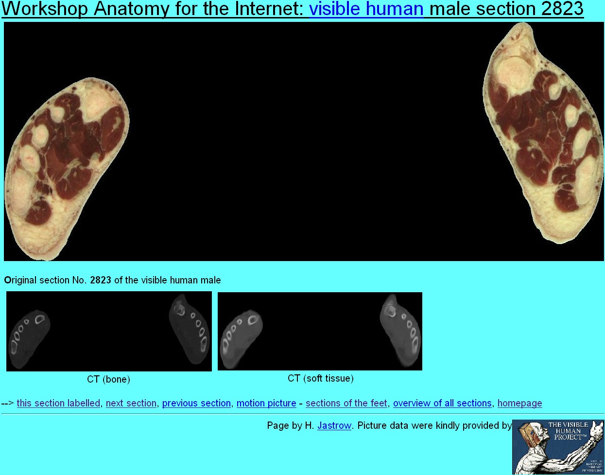
CT-Scans are shown below the original sections
for optimal space management in web pages with sections of the lower limb
(Fig.9).
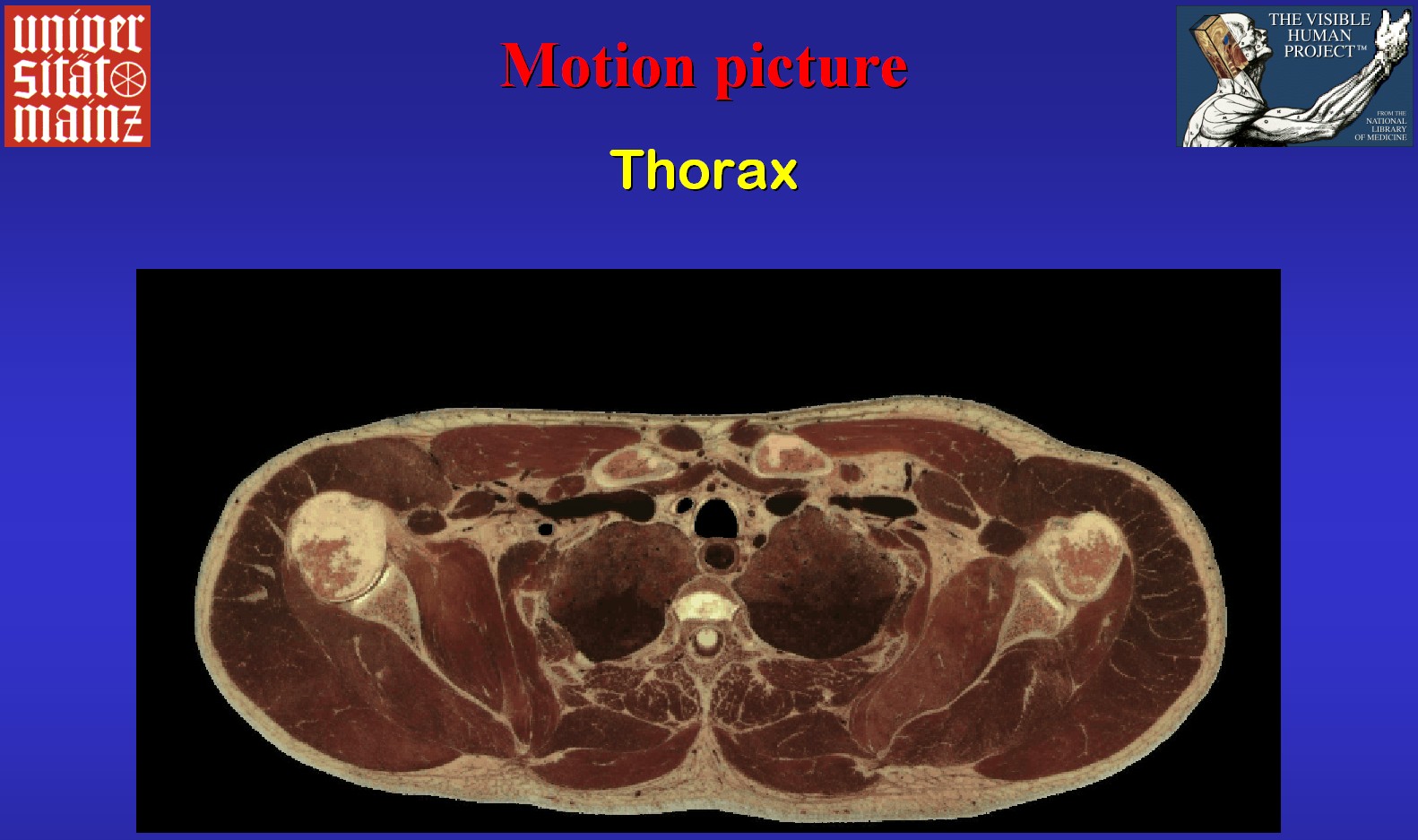
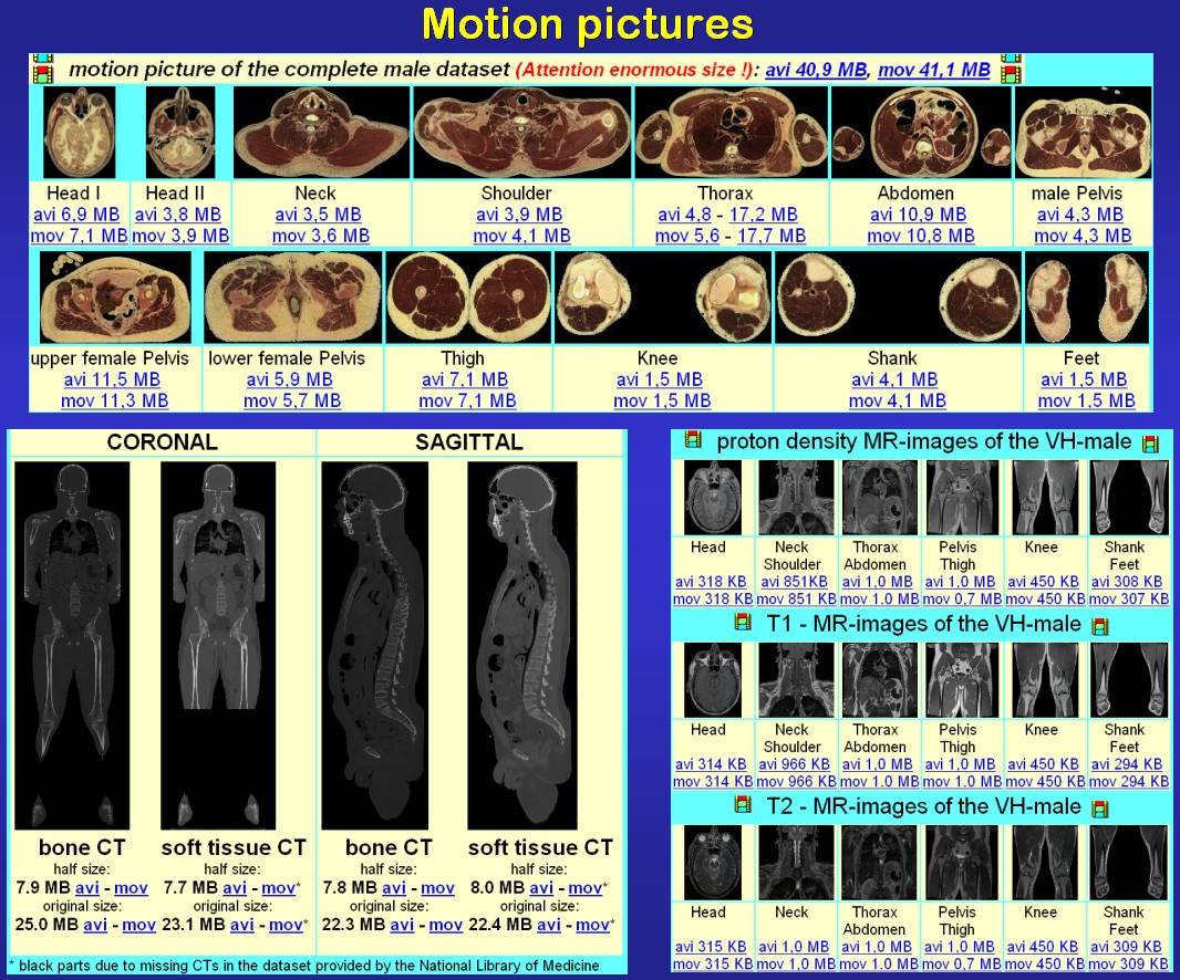
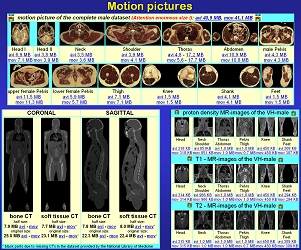
The motion pictures of the web atlas were created
from stacks of trimmed, aligned, cleaned and equally sized images of all
the anatomical or radiological sections mainly of the VH-male provided
by the NLM (Fig.10). They were produced using Ulead Media Studio
or MainActor software in either AVI or Quicktime format, both in Cinepack
Radius compression. Only some of the motion pictures of the original sections
are reduced in size due to the enormous amount of data involved. One motion
picture even runs through the scaled down total of the complete sections
of the VH-male. |
Fig.10 (link to atlas motion picture
[17 MB!])
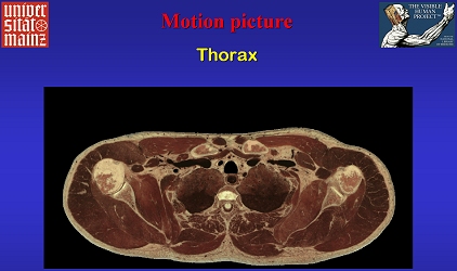 |
Fig.11 (link to atlas page)
 |
In Fig.11 you see three of five pages
providing a total of over 120 motion pictures ready for download that were
mainly produced from the VH-male data set.
The atlas of visible human sections is one of
some anatomy web-teaching modules of the Workshop
Anatomy for the Internet of the Johannes Gutenberg University of Mainz,
Germany. These modules are linked to each other as shown in Fig.12. |
Fig.12
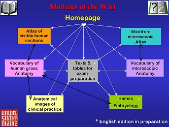 |
Fig.13 (link to atlas homepage)
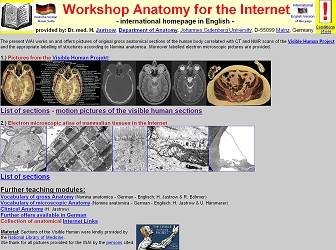 |
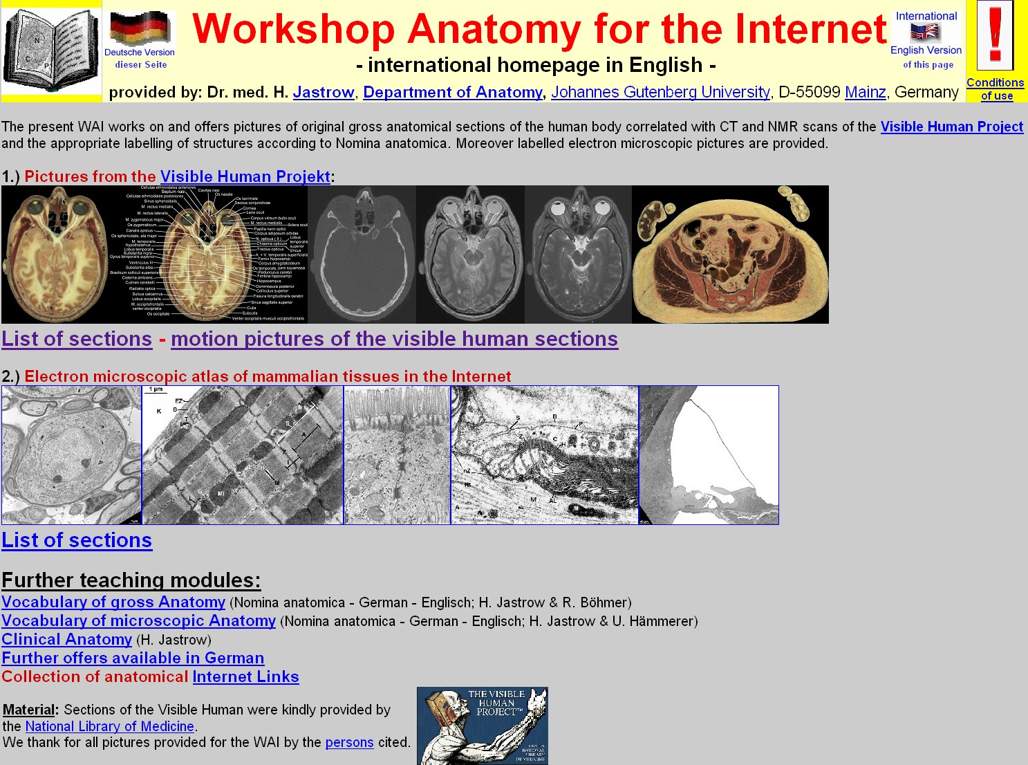
Fig.13 shows the English
homepage of the Workshop. Apart from the extensive
electron microscopic atlas and the other teaching modules it leads
to the atlas of visible human
sections in the internet which is presented here. When choosing List
of sections, the index page of
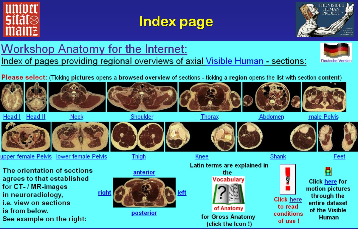
this atlas (Fig.14) is loaded.
This index page provides miniaturised transverse
overview sections for each region of the body, linked to pages with browsed
overviews of the chosen sections. Further one section is labelled with
terms of orientation providing a link to an appropriate page of a vocabulary.
Additional icons provide links to this vocabulary
of gross anatomy, motion pictures,
language selection and conditions of use.
If you click on the head 1 icon you will reach the first region overview
of the head. |
Fig.14 (link to atlas index page)
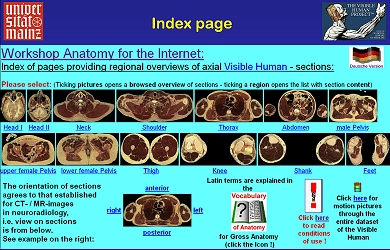 |
Fig.15 (link to atlas page)
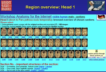 |
The first
region overview of the head providing small icons of all sections chosen
here (Fig.15). When you click an icon the page with the full sized section
and its corresponding radiological images, in the head region MR and bone
CT come up. A tick on the section number below opens the page with this
section labelled. Further down you see the beginning of the list of the
most important structures of the sections.
The linked vocabulary
of gross anatomy (Fig. 16) provides over 850 Latin anatomical
terms and their corresponding expressions in English and German. The terms
are arranged according to subject as well as alphabetically. Thus unclear
labelling can be looked up easily. |
Fig.16 (link to atlas page)
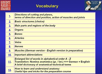 |
Fig.17
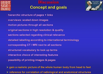 |
By the points listed in Fig.17 the atlas
intends to be a useful online reference for medical education and research.
It enables a visual journey through the whole body showing all relevant
structures of gross anatomy and in so far contributes to a three-dimensional
understanding of topographic relationships. All important structures of
gross anatomy are labelled using the official terminology to make the atlas
ideal for use throughout the world. By giving representative views of all
relevant structures of all body regions next to corresponding radiological
images a valuable reference for correlation of radiological and anatomical
structures has been created.
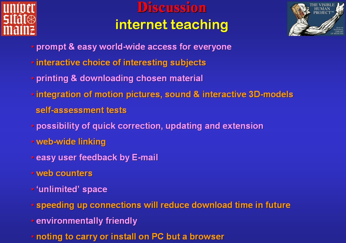
In Fig.18 you see why the present atlas
is published the internet as the most important medium of today and future
times to retrieve and present knowledge. Namely, the web offers possibilities
that partly even CDs cannot provide and that are of great advantage in
comparison to printed media. Since it is the aim to offer entirely correct
labelling, the users of the atlas are requested to inform the author of
imperfections via E-mail, as, in
contrast to a book, publication in the internet offers the possibility
of quick and easy correction. |
Fig.18
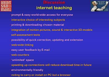 |
Fig.19
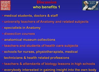 |
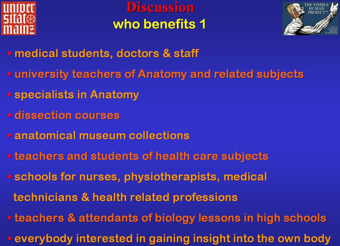
Many people can benefit from the internet atlas
since the offered material can be used for teaching as well as learning
gross anatomy in general (Fig.19). The spectrum reaches from university,
health care related schools and colleges to lay people interested in the
construction of the human body.
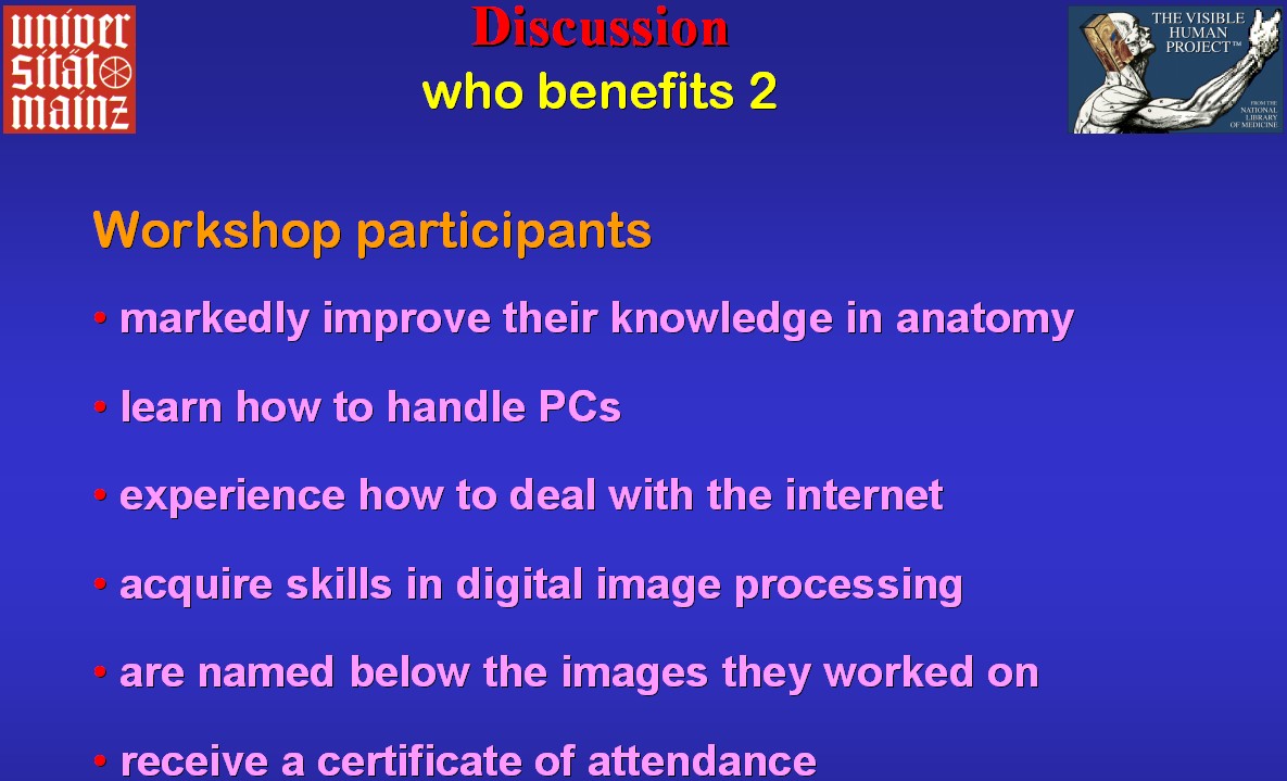
The students who help to create teaching material
for others by labelling the images in the workshop benefit in many ways
(Fig.20) and acquire a special qualification useful for their later
work as medical doctors and when seeking positions. |
Fig.20
 |
Fig.21
 |
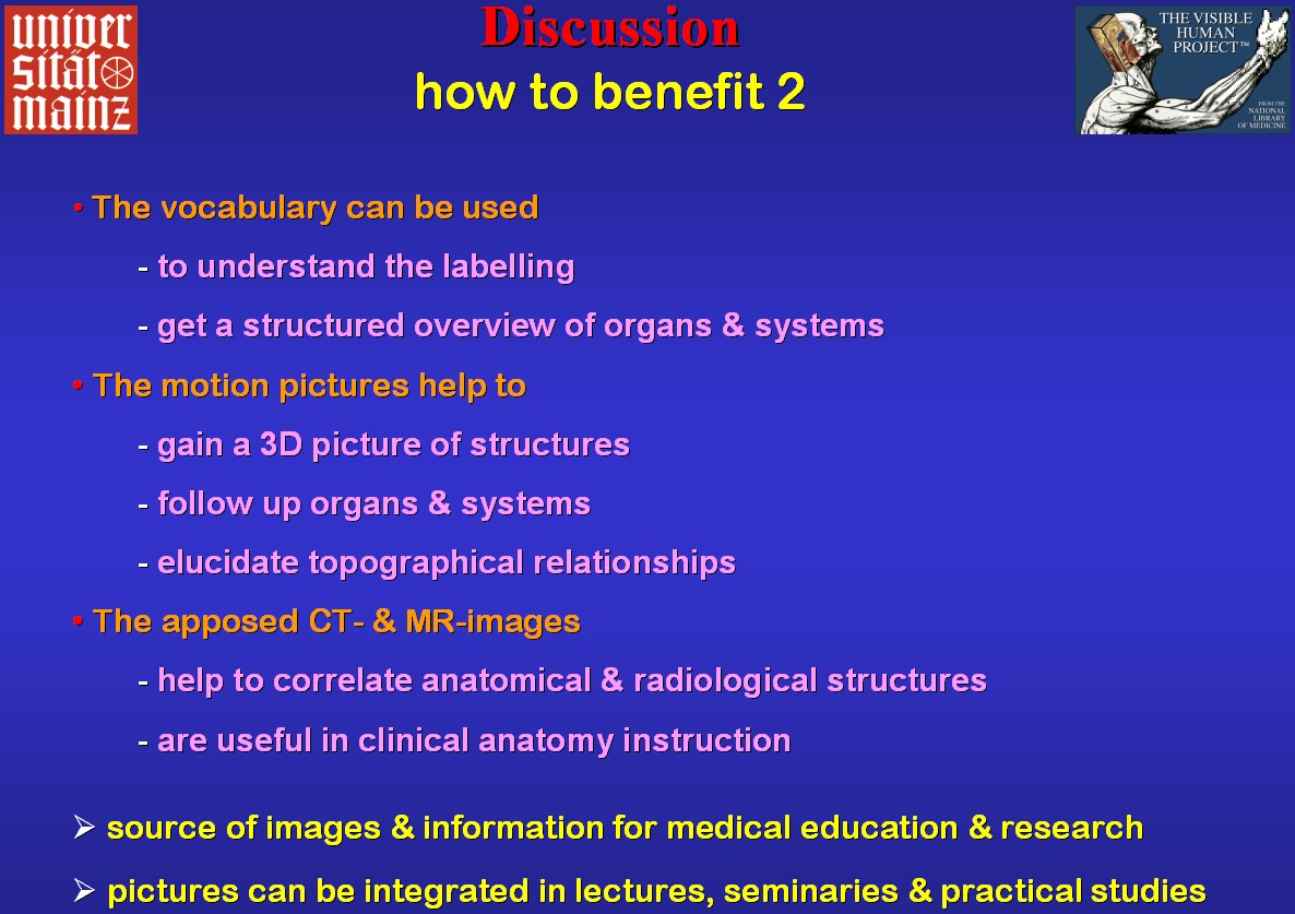
The offered material is suitable for study and
instruction of anatomy in general. In Figs.21 & 22 you
see some examples what it can be used for. The broad spectrum of usability
spans from university instruction of specialists and reference for scientific
purposes over education of medical students, doctors and staff, teachings
of clinical anatomy to biology lessons in college and high school. Either
direct internet access or printouts of the images are applicable. For non-professionals
the vocabulary is a valuable key to understanding the international terminology.
The sequence of sections and motion pictures facilitate acquisition of
a 3D-understanding of anatomy and topographical relationships. In the training
of medical students and doctors it is essential to correlate anatomical
structures of the human body with radiological images. For this reason
sections are presented next to corresponding radiological images that also
can serve radiologists as a reference. In brief the atlas is a source for
many uses. |
Fig.22
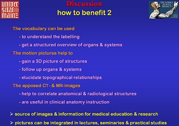 |
Fig.23
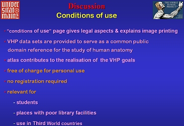 |
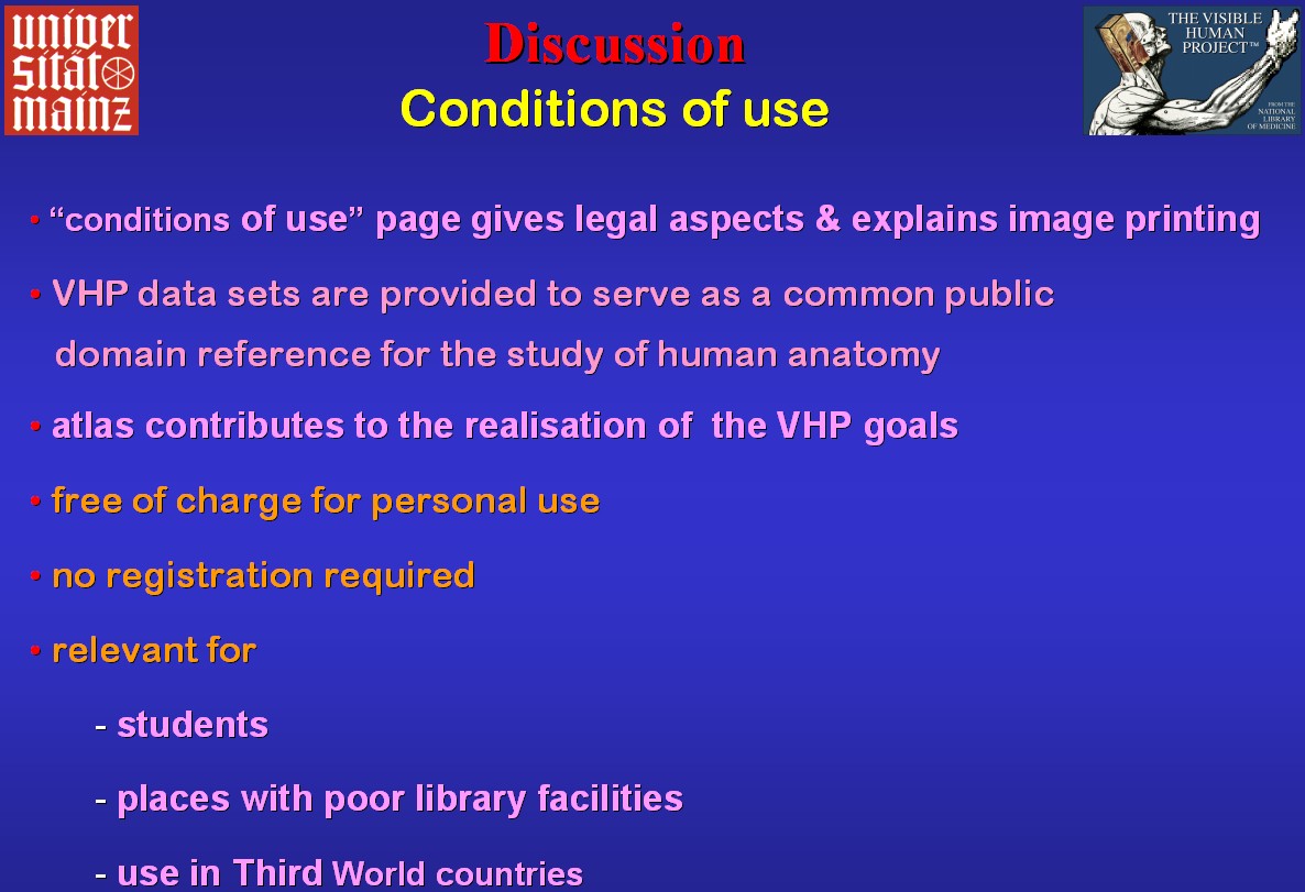
A page with the conditions
of use informs about legal aspects and explains how to obtain printouts
of the images. High quality anatomy software is often not affordable for
students. In contrast, the present offer, even though no freeware, allows
everybody to benefit from the excellent material offered by the NLM. In
so far it contributes to the realisation of the goals of the Visible Human
Project (Ackerman 1998) by being easily accessible in the Internet and
at no charge for personal use (Fig.23).
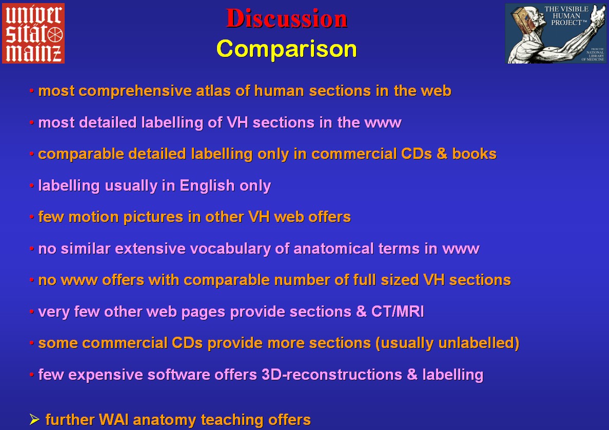
It is apparent that the present atlas is the most
comprehensive one in the internet offering most detailed labelling of sections
in the web. Otherwise, comparable detailed labelling, usually in English,
is only present in few commercial CDs and books. For the reasons listed
in Fig.24, the present contribution surveys other web-anatomy atlas
projects and commercially available software. With its further anatomy
teaching offers the Workshop Anatomy for
the Internet represents a unique concept of presenting anatomy to specialists
as well as to the general public. |
Fig.24
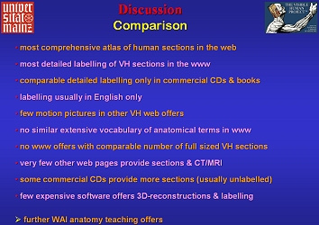 |
Fig.25
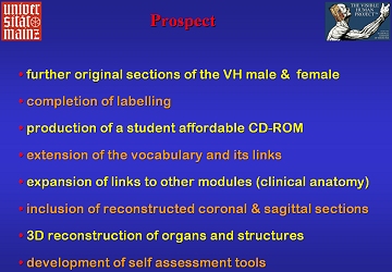 |
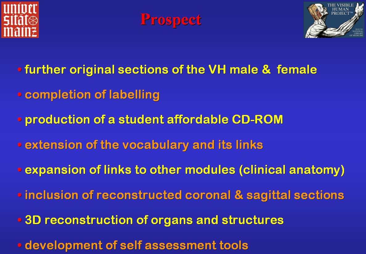
The present atlas is constantly extended by including
further original and labelled sections aiming at a maximal intersection
distance of 5 millimetres in all regions (Fig.25). Due to considerable
download time, especially for the motion pictures, it is planned to offer
them on a student affordable CD-ROM with as many sections as possible.
Apart from the extension of the vocabulary it is planned to offer computed
coronal and sagittal sections, 3D reconstructions and self assessment tools.
With these goals it is intended to contribute to world-wide improvement
of 3D-understanding of anatomy in pre-clinical and clinical medicine.
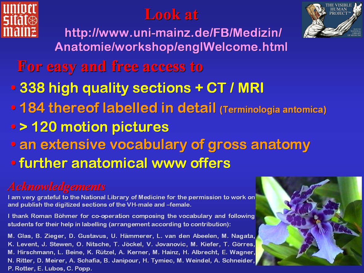
With the summary of Fig.26 I want to invite
you to explore the internet pages of the Workshop
Anatomy for the Internet. Further, I acknowledge those who supported
this project. |
Fig.26
 |

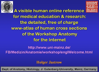
 A
visible human online reference for medical education & reseach
A
visible human online reference for medical education & reseach