Overview macrophages (Macrophagocyti):
Pages with explanations are linked to the
text below the images if available! (Labelling is in German)
 |
 |
 |
 |
 |
 |
 |
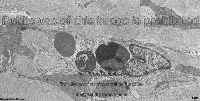
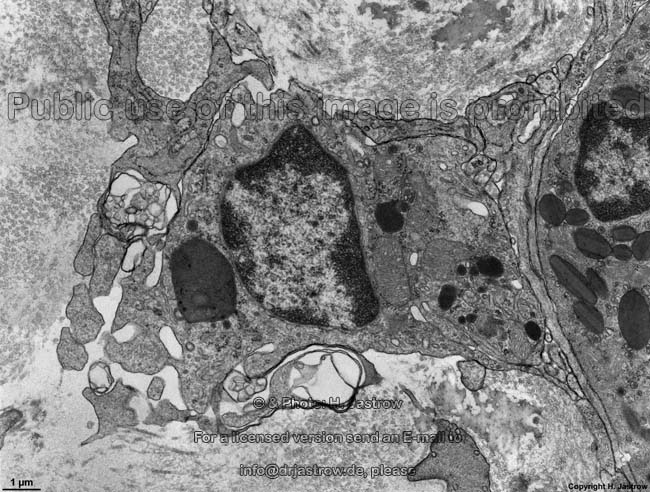
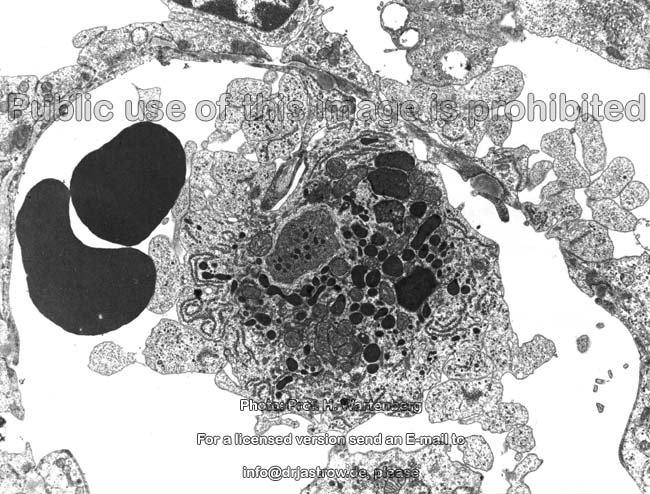
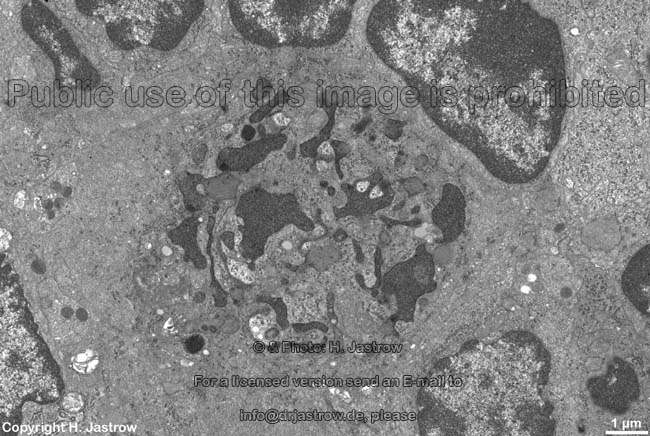
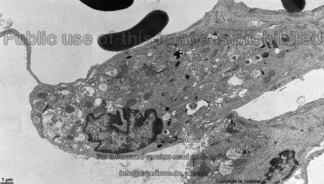
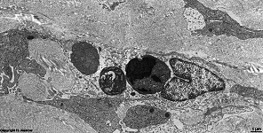
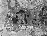
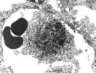
macrophage with large phagolysosome in con-
nective tissue of lamina propria in stomach (rat) |
detail thereof:
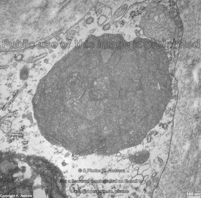
phagolysosome 1 |
detail 2
phagolysosome 2 |
detail 2
phagolysosome 3 |
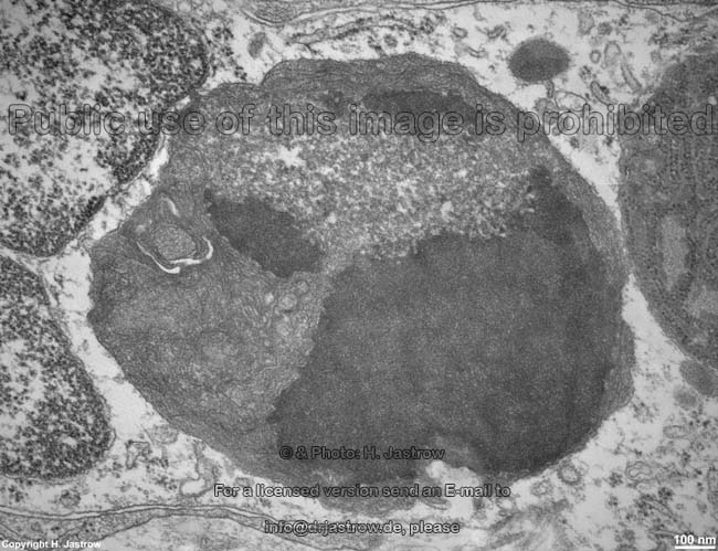
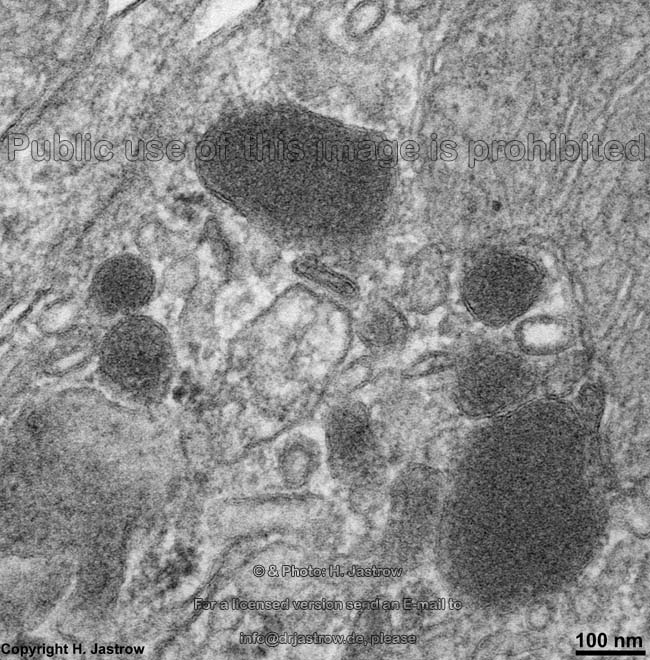
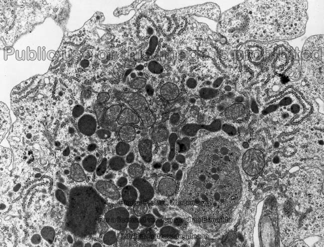
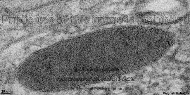
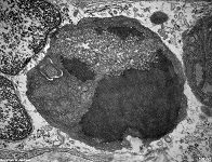
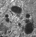
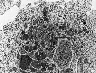
macrophage, Tela
submucosa gastrici (rat) |
detail 1: primary
lysosomes |
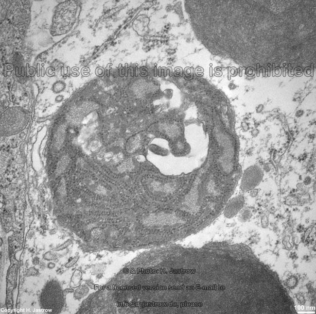
detail 2: hetero-
lysosome |
 |
 |
 |
 |
 |
 |
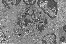 |
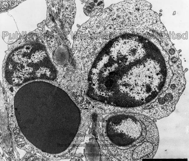
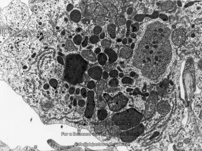
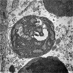
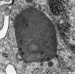
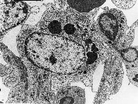
macrophage of the
spleen 1 (monkey) |
macrophage of the
spleen 2 (monkey) |
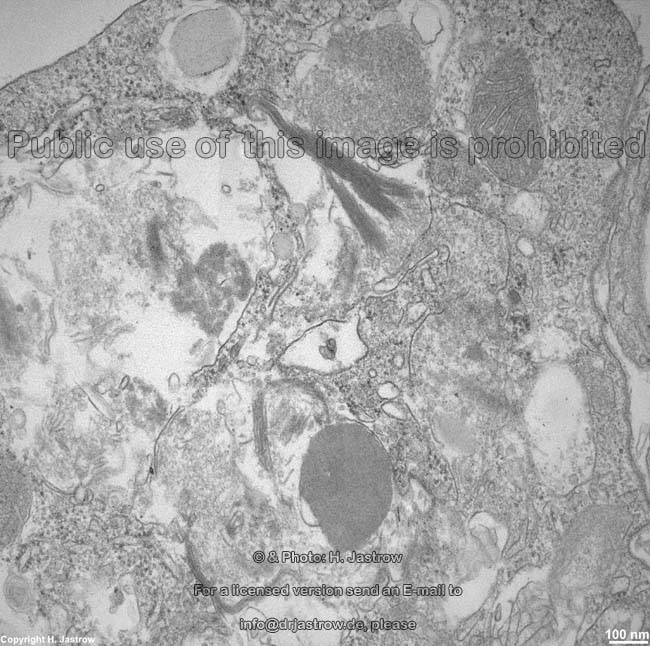
detail 1 of macrophage
spleen 2 (monkey) |
detail 2 of macrophage
spleen 2 (monkey) |
macrophage of the
spleen 3 (monkey) |
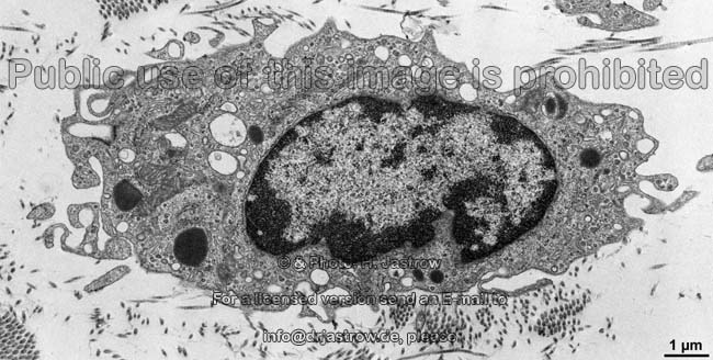
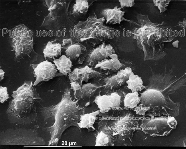
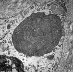
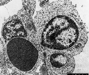
macrophage in loose connective tissue
= histiocyte (rat) |
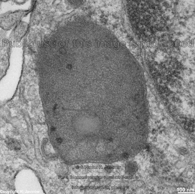
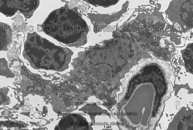
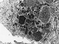
macrophage with hetero-
lysosomes spleen (rat) |
Macrophages (Terminologia histologica: Macrophagocyti)
are cells that "eat" extracellular particles. They are of
importance for defense of bacteria and remove foreign bodies,
substances and molecules. Further these cells release several factors
which influence other cells of the immune system e.g., interleukin-1
which activates and attracts neutrophils.
They further serve for destruction of abnormal cells (tumor,
virus infected or overaged cells) e.g., in spleen
for elimination of overaged red blood cells which
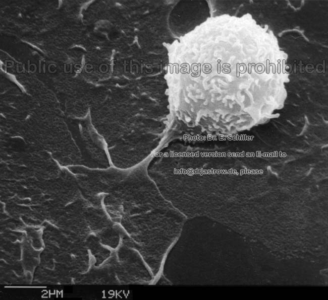
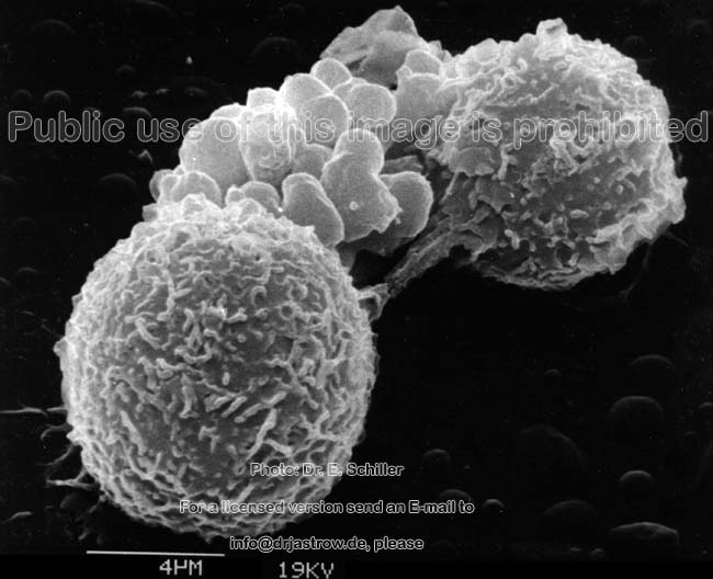
are poor in distorsion. The macrophages grasp particles with their mobile
processes (pseudopods) and draw them into
their interior whereby the get a limiting membrane deriving form the cell
membrane. This procedure is called phagocytosis.
Incorporated particles are called phagosomes
or endosomes or in case they have more than one layer of outer membrane
coat phagophores. Lysosomes
fuse with the endosomes to form heterolysosomes
= phagolysosomes and their enzymes begin to destruct the content. Phagocytosis
is evoked when surface antigens of particles (e.g., viral
coat proteins) get in contact with the cell
membrane of pseudopods of macrophages.
By their mobility one can distinguish either resting
macrophages (Terminologia histologica: Macrophagocyti sessiles) or wandering
macrophages also called histiocytes (Terminologia histologica: Macrophagocyti
mobiles).
Macrophages are also essential for getting rid of degenerating or
insufficiently working cells of the body e.g, in spleen
where they invaginate and phagocyte
old inflexible erythrocytes or they serve to
eliminate virus infected cells.
Special types of macrophages that
are restricted to defined locations are the microglia
cells of the central nervous system,
the Kupffer cells attached to
the endothelium of the sinusoids
of the liver, the alveolar
macrophages inside the cavities of the alveols
of the lung in order to phagocyte
dust particles, the previously mentioned phagocyting
reticulum cells in the red pulp
of the spleen and the A-synovialozytes
located in the synovia of joints (synovial membrane) as well as the peritoneal
macrophages patrolling the surface of the peritoneum.
All these cells belong to the mononuclear
phagocyte system.
--> connective tissue, free
cells in connective tissue, blood cells,
mast
cells, plasma cells, pseudopods,
lysosomes,
heterolysosomes,
phagocytosis
--> Electron microscopic atlas Overview
--> Homepage of the workshop
Some images were kindly provided by Dr. E. Schiller
or Prof. H. Wartenberg; other images, page & copyright H. Jastrow.