Overview mast cells (Mastocyti):
Pages with explanations are linked to the
text below the images if available!
 |
 |
 |
 |
 |
 |
|
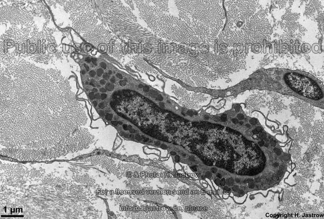
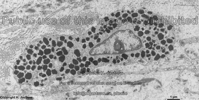
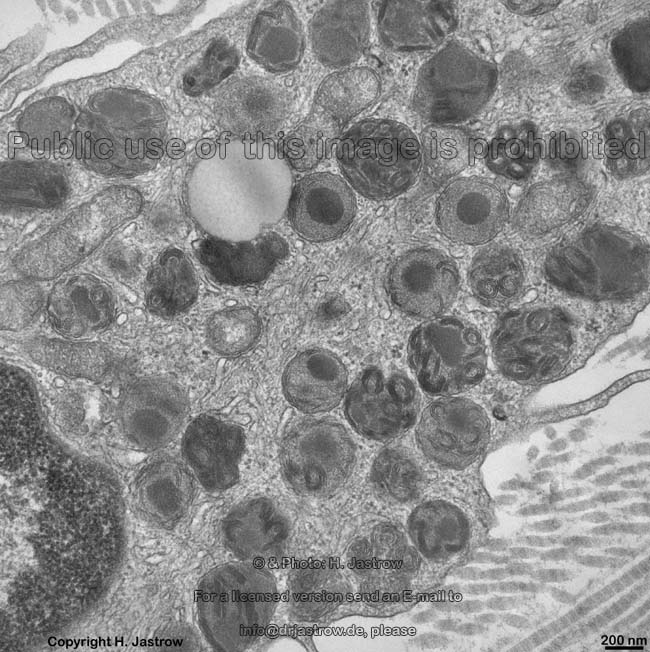
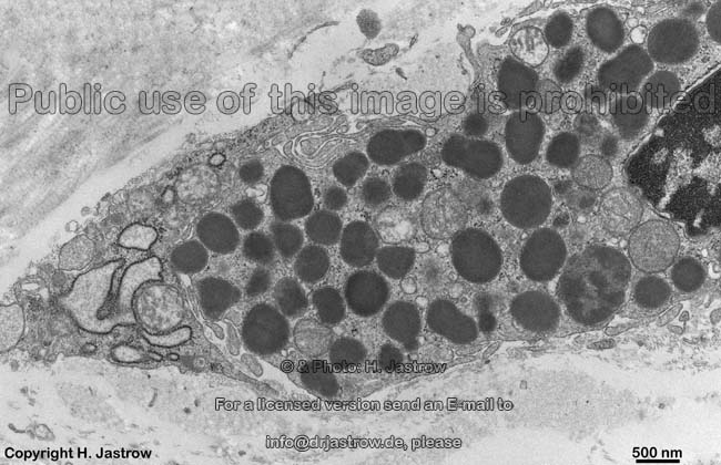
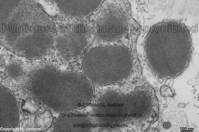
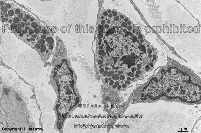
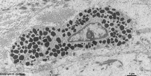
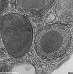
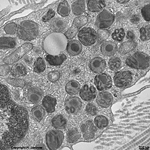
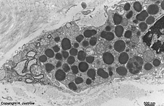
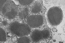
human mast cell (type 1)
from subcutis |
left detail thereof 1 |
right detail thereof 2 |
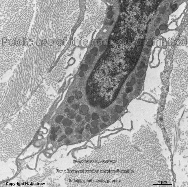
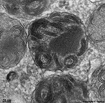
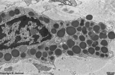
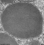
human mast cell
(type 1) |
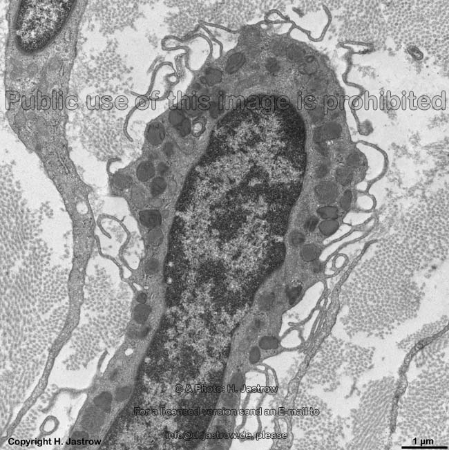
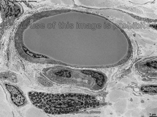
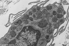
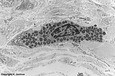
human mast cell (type 1) from the sclera of
the eye-bulb (4 hrs post mortem fixed) |
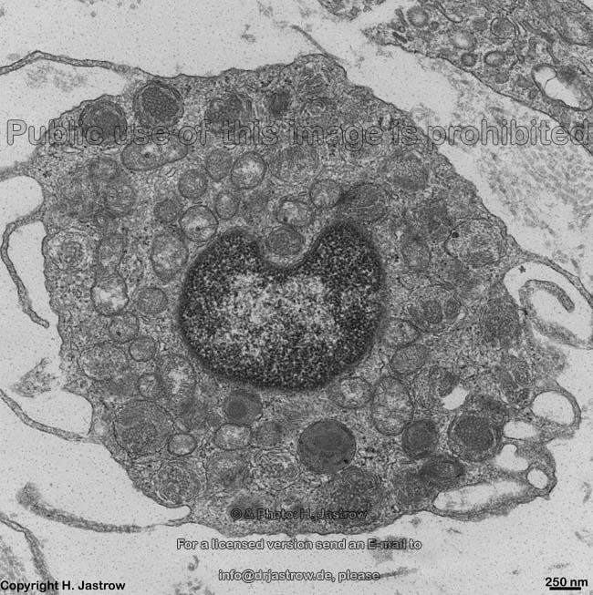
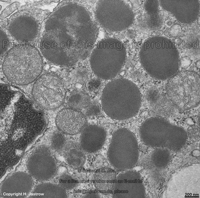
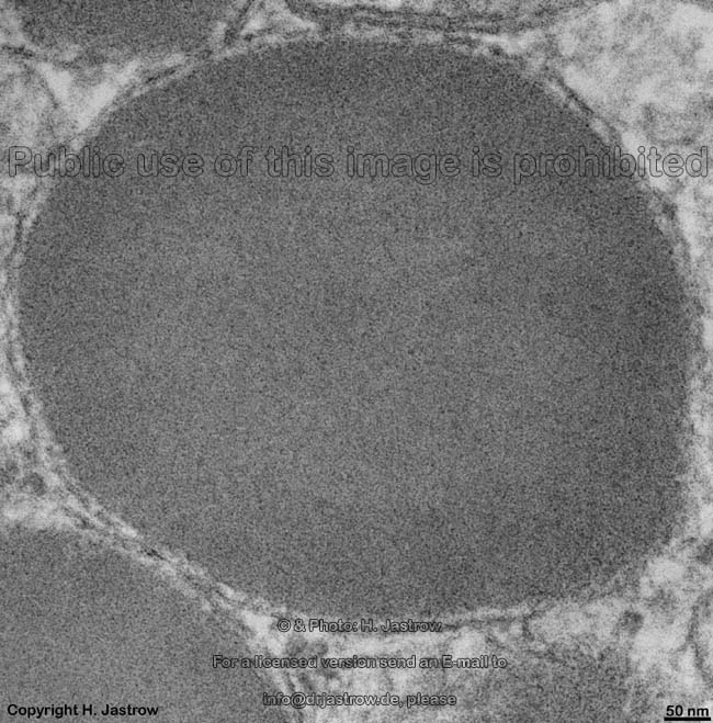
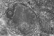
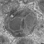
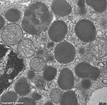
human mast cell
vesicles 1 |
 |
 |
 |
 |
 |
human mast cell
(Type 1)
|
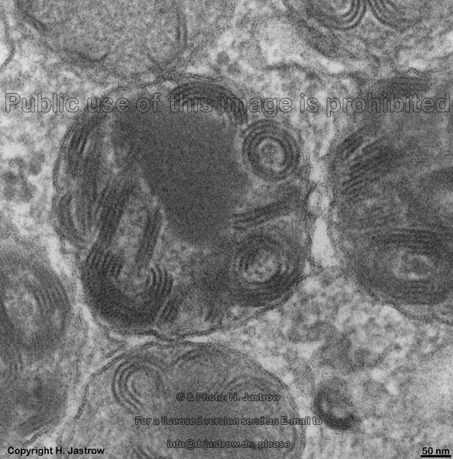
human mast cell
vesicles 2 |
human mast cell
vesicles 3 |
human mast cell
vesicles 4 |
human mast cell
vesicles 5 |
 |
 |
 |
 |
 |
 |
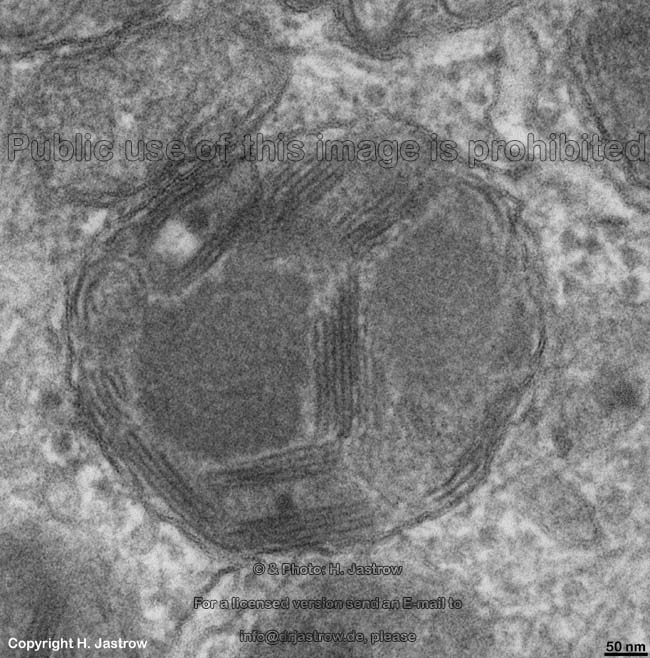
human mast cell
(type 1)
|
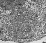
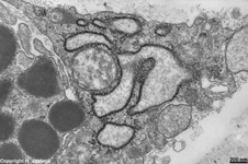
human mast cell vesicles some of
which are in state of regeneration
|
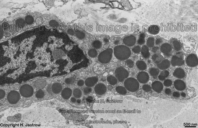
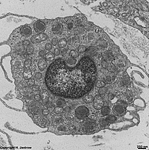
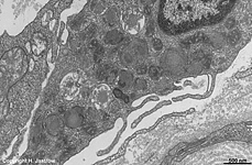
human mast cell
(type 2)
|
detail: human mast
cell vesicles 6
|
detail: human mast
cell vesicles 7
|
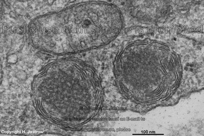
human mast cell vesicles 8
from a further mast cell
|
 |
 |
 |
 |
 |
|
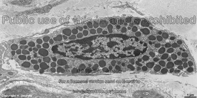
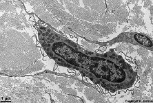
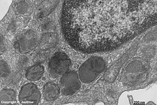
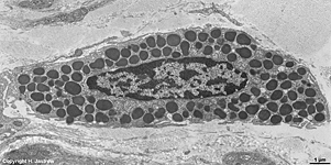
rat mast cell 1
|
anterior detail thereof |
posterior detail thereof |
rat mast cell vesicles 1 |
rat mast cell vesicles 2 |
 |
 |
 |
 |
 |
rat mast cell vesicles 3 and an
undamaged extracellular vesicle
|
rat mast cell
vesicles 4 |
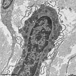
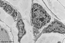
univacuolar fat cell
+ rat mast cell 2 |
previous mast cell of the rat in detail |
mast cells of rat 3 |
A mast cell (Mastocyte; Termionologia histologica: Mastocytus)
is a free cell of the connective
tissue. According to their location mast cells on their most frequent
places have a special terminology: either as mucosa related mast cells
(Termionologia histologica: Mastocyti mucosales) or as perivascular mast
cells (Termionologia histologica: Mastocyti perivasculares). Mast cells
are actively mobile, i.e. they are able to migrate through surrounding
extracellular matrix using their pseudopods
(or filopods) which are several micrometers long and motile. The mast cell
cytoplasm
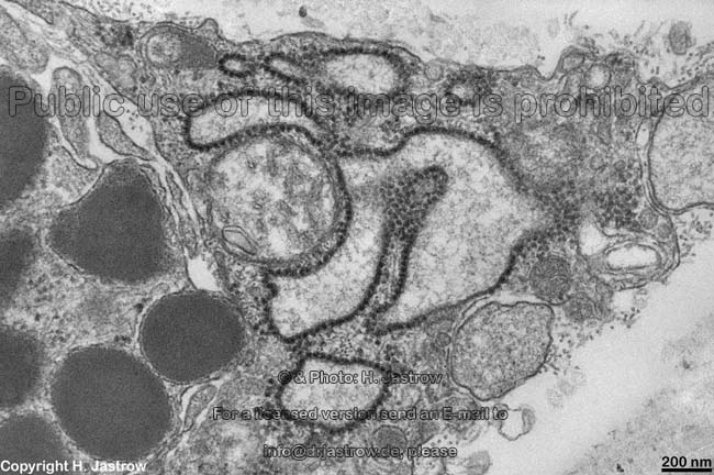
contains several hundred vesicles (NO granules!). Stimulation by
Immunglobulin
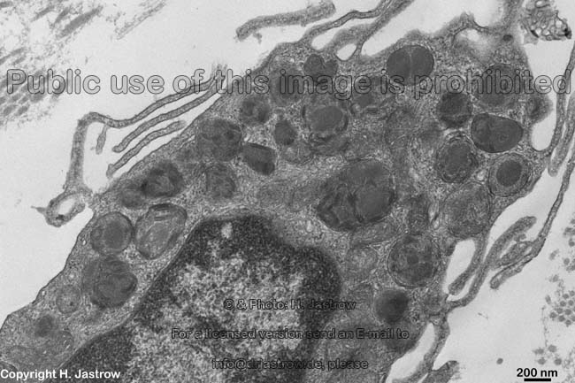
E (IgE) effects on one hand an exocytosis: vesicle fusion with the
cell membrane and release of vesicle
content on the other hand some intact vesicles (with membrane) leave the
cell (image on the lower left). Fast release of vesicle content is of major
imoprtance in allergic reaction of the immediate type (Type 1 allergy).
After a necessary first contact with the antigen specific IgE antibodies
are produced by
plasma cells, these IgEs
bind on mast cells that then begin to produce an FceRI receptor which is
expressed on the cell membrane. In case
of new presence of the antigene the latter binds to the FceRI receptor
of the sensitized mast cell and causes the allergic reaction proviously
mentioned. This immediate release of mast cell vesicle content has been
calledcompound exocytosis. The
expression mast cell degranulation meaning the same is based on early light
microscopy and is not correct since infact it is a fast exocytosis of vesicles
characterized by a surrounding membrane. Accordingly, the established term
mast cell granule is not correct since a true granule like e.g., a Glycogen
granule is not surrounded by a membrane. As can be seen in the images,
mast cell vesicles of the rat are very electron-dense and homogenous.
In Man different kinds of vesicles are encountered. The maximal
diameter of the vesicles is about 1 µm. They
are basophilic, water soluble and metachromatic in some light
microscopic stains.
Chemically the vesicles contain water, histamin,
heparin and glykosaminoglycans (consisting of glucoronic acid, glucosamin
and sulphuric acid, explaining metachromasy), chemotactic factors, tryptase
and/or chymase. In rodents like rat further serotonin. Further, mast cells
release mediators, of which in vesicles are stored: tumor-necrosis
faktor alpha (TNFa), interleukin 4,5,6 and 8
(IL4, IL5, IL6,IL8) as well as eosinophil chemotactic factor stimulating
immigration of eosinophilic granulocytes which then bloc histamine effects
and help in limiting an inflammation. In addition the mast cell vesicles
contain bFGF, stem cell factor, endothelial permeability factor and vascular
endothelial growth factor. Further mediators are newly synthetised in mast
cells only when stimulated and immediately released. Therefore they are
probably not stored in the vesicles: prostaglandin GD2 (PGD2), Leukotrien
C4 (LTC4), Leukotrien D4 (LTD4), platelet activating factor (PAF).
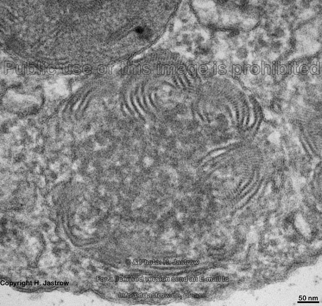
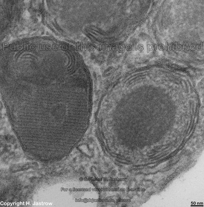
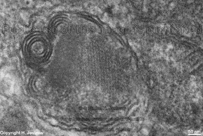
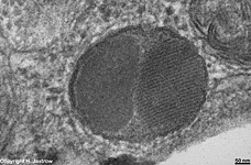
Electron microscopic aspect of mast cell vesicles: there are
different components in human cylindrical membrane scrolls (see
image of mast cell vesicles 6), crystals (notable on the regular
striping; see image of mast cell vesicles 2,3,5,7). Some authors even clain
the presence of ribosomes inside mast cell
vesicles.
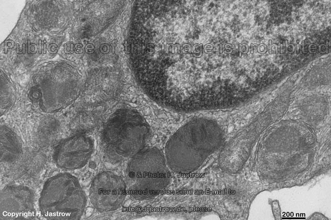
Usually 7,000 to 10,000 mast cells are present in 1
mm³ of the skin mostly close to capillaries,
lymph
vessels and nerves. Human mast cells are
usually elongated cells about 5 x 15 µm in size. They derive
from a multipotent stem cell in bone marrow
that expresses on its surface, i.e. on the cell membrane a CD34 receptor.
These stem cells may also develop into basophilic
granulocytes. The direct precursors of the mast cells have no vesicles
and look like monocytes. They are distributed
by the blood and emerge into connective tissue
on any location. Some hours later they develop fist vesicles and become
mature mast cells. Basing on morphology and immunhistochemistry 3 types
of mast cells are distinguished:
Type 1 most common, predominant in skin,
mainly contain vesicles with amorphic electron dense content. In some cases
scroll-like electron dense structures are located at the side of a partly
cristalloid vesicle core (detail images 1-5). This type of mast cells synthetises
tryptase and chymase.
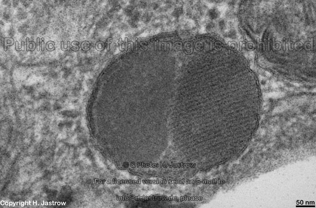
Type 2 less common, predominant in lung,
show mainly vesicles with scroll-like content and osmiophilic lipid
droplets of varying electron density (detail images 6-8). The lipid
bodies contain cyclooxygenase. The mast cells of type 2 only contain tryptase.
Type 3 these postulated rare type only synthetises chymase.
It shall be present in axillary lymph nodes, the
lung
and in intestinal connective tissue.
Mast cells show only small amounts of rough endoplasmic
reticulum and small Golgi fields. Besides
the vesicles their cytoplasm
contains some free ribosomes, microtubules,
actin-
and intermediate filaments. Lipid
bodies are only present in type 2 mast cells. Besides the fast IgE
stimulated exocytosis of vesicles,
those may also be released much slower by other substances; this procedure
is termed peacemeal degranulation.
Mast cells are involved in allergy, acute and chronic inflammation
processes, T-cell activation and tissue defense
of parasites. Their function is improper in many deseases of the skin
e.g., psoriasis, chronic eczema, sclerodermia, lichen simplex and - planus.
--> connective tissue, free
cells in connective tissue, plasma cells,
lymphocytes,
blood
cells,
basophilic granulocytes, bone
marrow
--> Electron microscopic atlas Overview
--> Homepage of the workshop
Images, page & copyright H. Jastrow.