Overview periphral nerve tissue
(Nervi
peripherici):
Pages with explanations are linked to the
text below the images if available! (Labelling is in German)
|
|
|
|
|
|
|
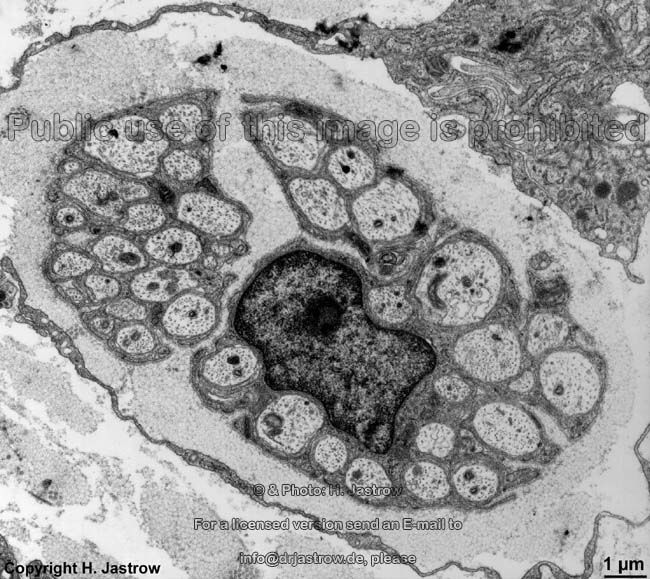
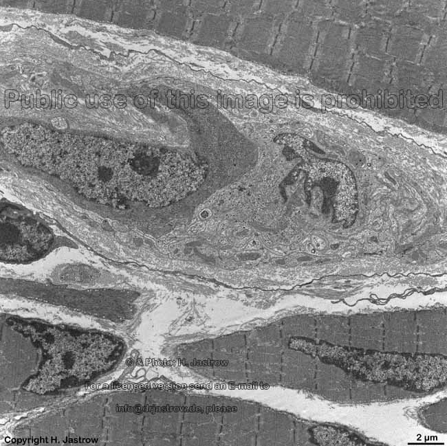
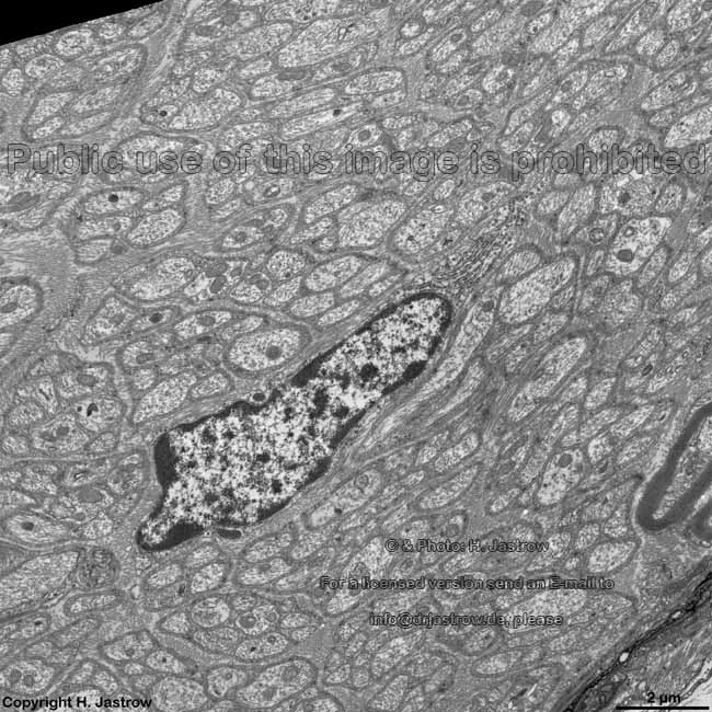
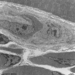
Plexus myentericus,
ganglion cell (rat) |
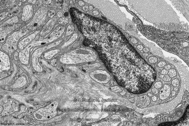
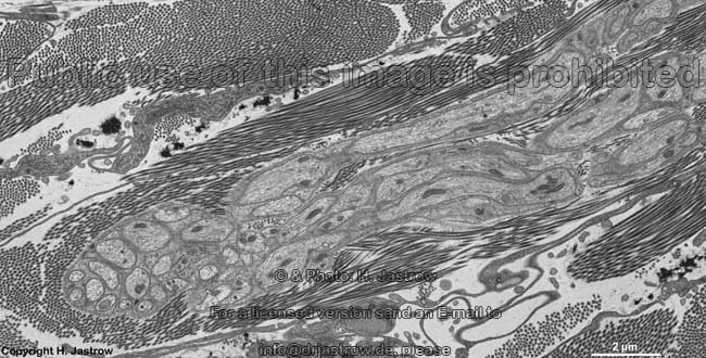
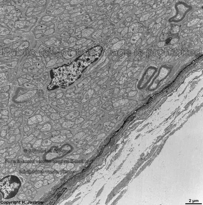
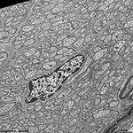
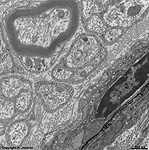
part of the nervus
vagus (rat) |
detail thereof: mainly
non-myelinated fibres |
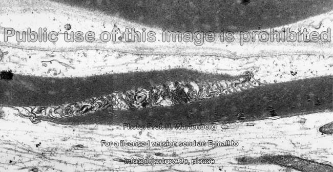
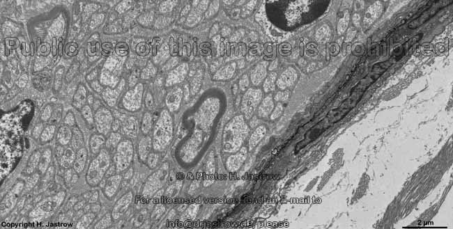
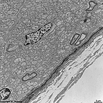
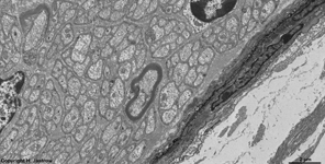
epineurium of the nervus vagus
in the adventitia of esophagus (rat) |
epineurium vagus
nerve (rat) |
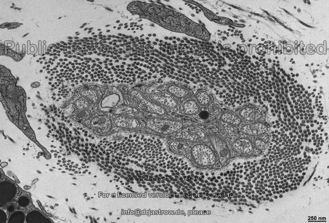
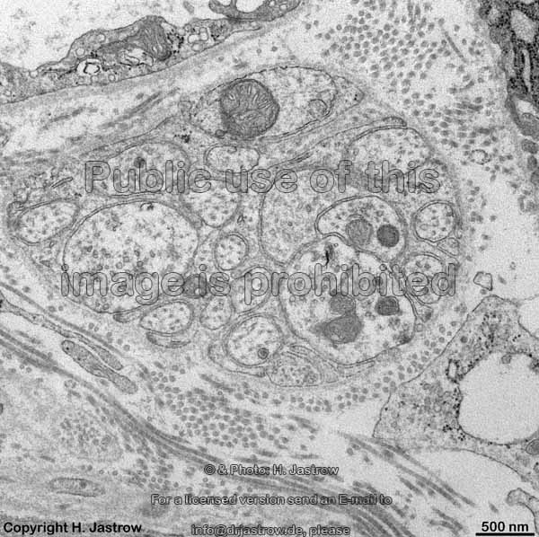
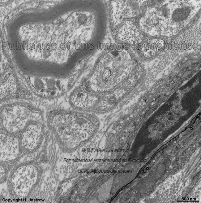
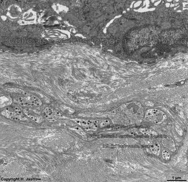
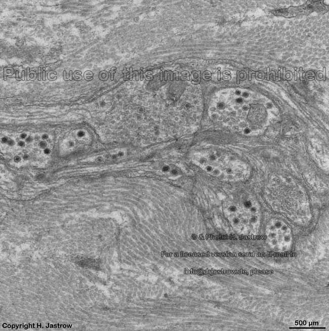
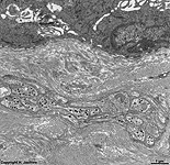
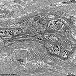
non meylinated nerves of the
Plexus submucosus (rat) |
detail thereof in L.
propria mucosae |
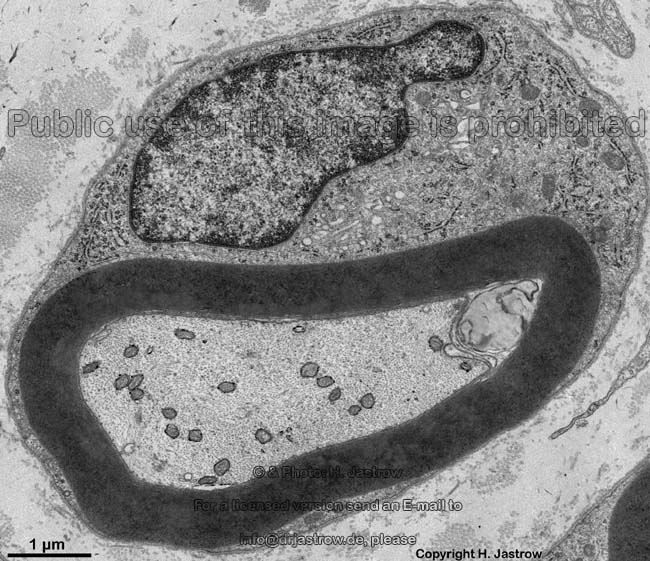
A nerve (Terminologia histologica:
Nervus) thus is defined as a bundle of processes of nerve cells,
here peripheral nerve fibres (Terminologia histologica: Neurofibrae periphericae)
with the surrounding layers of connective tissue.
In case of larger nerves when looking from outside to inside we
can distinguish the following
layers:
1. The epineurium (Terminologia
histologica: Epineurium) ensheats the entire nerve and is in continuity
with the dura mater (pachymeninx; Terminologia
histologica: Dura mater, Pachymeninx). It nourishes and protects
the nerve proper. The epineurium
has a superficial layer called superficial epineurium (Terminologia
histologica: Epineurium superficiale) consisting of irregular
woven connective tissue which is connected to surrounding connective
tissue and a deeper layer called deep
epineurium (Terminologia histologica: Epineurium profundum) of loose
connective tissue with intermingled unilocular
adipose cells, elastic networks
with arterioles and venules,
their supplying non-myelinated autonomic nerve
fibres as well as small vessels responsible
for blood supply. The elastic
networks allow a certain flexibility of the nerve and cause its retraction
in case of being cut. The epineurium covers
many fasciles (bundles of nerve fibres with some to some hundred single
fibres) and is in continuity with the connective tissue sheath of the
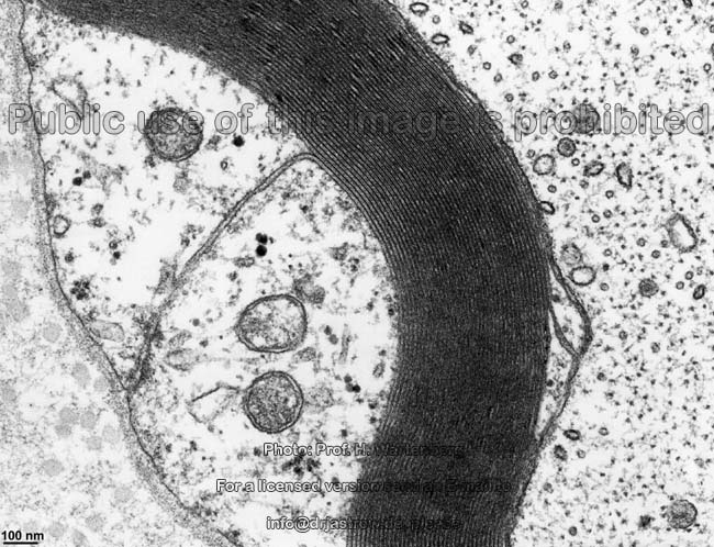
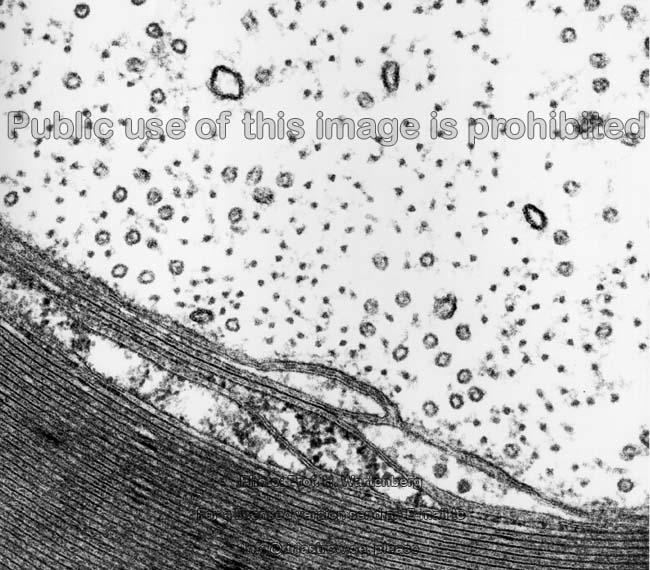
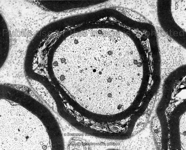
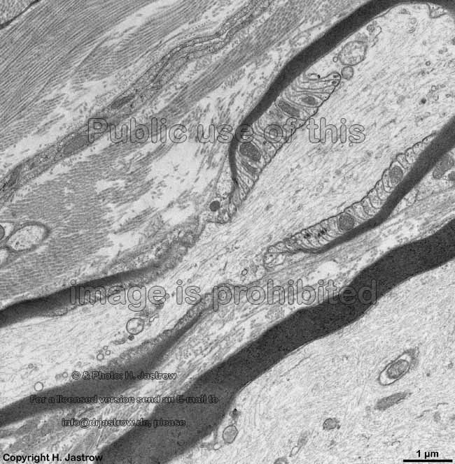
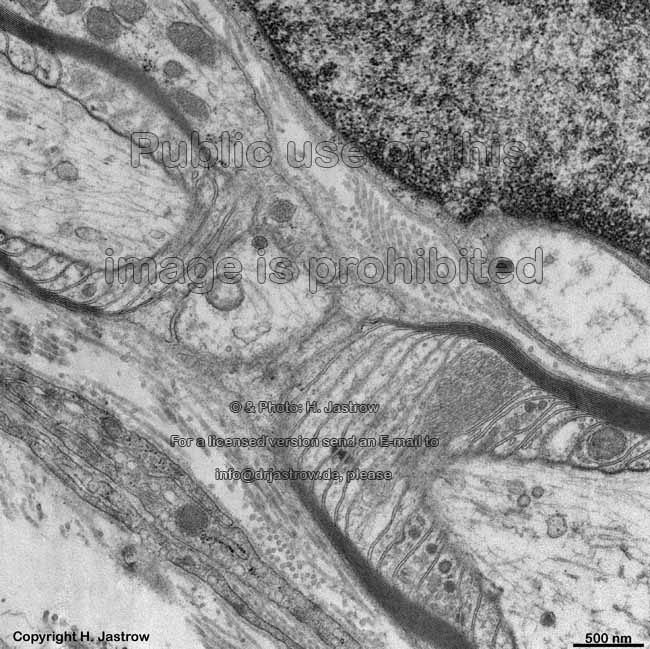
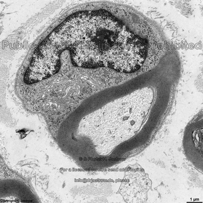
2. perineurium (Terminologia histologica:
Perineurium). This layer has an
outer
mechanically resistant layer of dense woven
connective tissue called fibrous part (Terminologia histologica:
Pars fibrosa) onto which the epithelioid
part, i.e. the perineural sheath (Terminologia histologica: Pars epitheloidea)
is attached. This layer consists of
up to 12 layers of very long, flat perineural cells (missing in
Terminologia histologica; proposal: Perineurocyti) which are surrounded
on all sides by basal laminas
and connected to each other by spot- and
belt
desmosomes as well as by tight junctions.
This is only the case in longitudinal direction and to the lateral edges
by not to covering or underlying cells which results in formation of tubular
sheath layers inside the epitheloid part. The cells are rich in caveols,
glucose transporter type 1 (Glut-1) and by
means of their tight junctions establish a
functionally relevant diffusion barrier for hydrophilic substances.
The 200 nm wide spaces between the cells is filled with collagen
fibrils mainly in parallel orientation and few elastic
fibres. The perineurium is maintained even in small branches of nerves
though it constantly reduces in in thickness until it finally gets lost
at the small terminal branches or it joins the capsular structures of specialised
nerve terminals like Meissner's corpuscules,
Vater-Pacini
bodies and others. The innermost layer which is located beyond the
perineurium is the
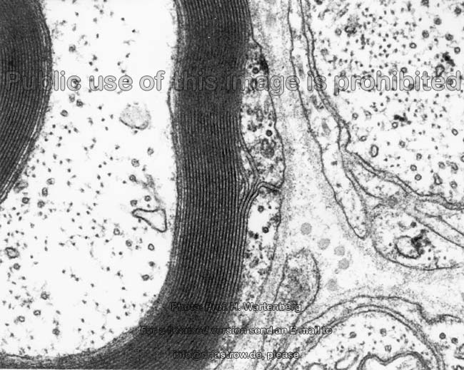
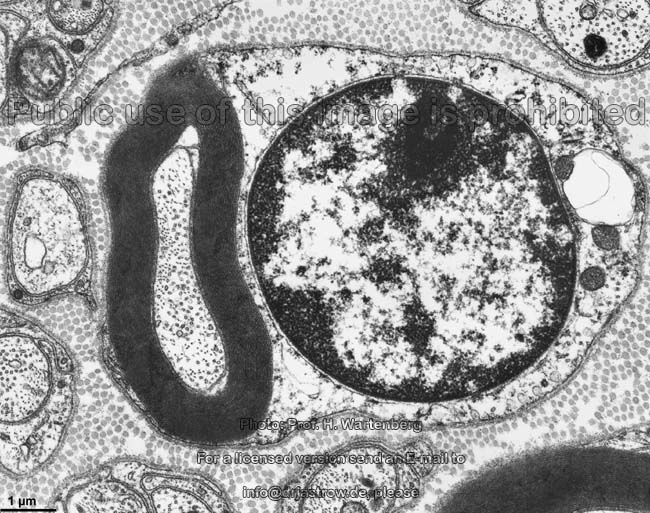
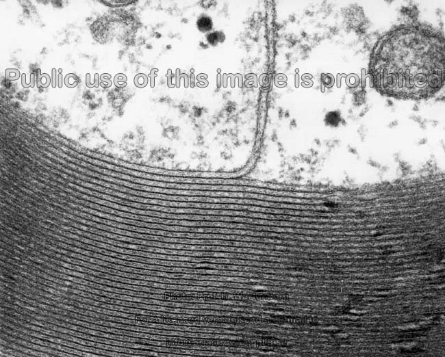
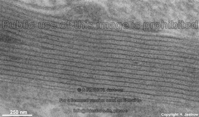
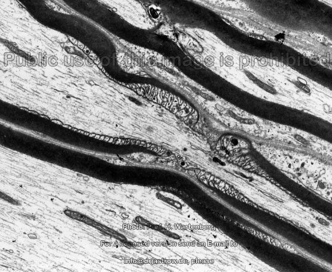
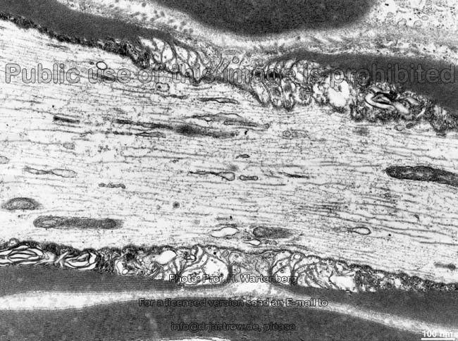
3. endoneurium (Terminologia histologica:
Endoneurium) which consists of loose
connective tissue. It fills the space between the single nerve fibres
which are enscheated by Schwann cells and
thus covered by a basal lamina.
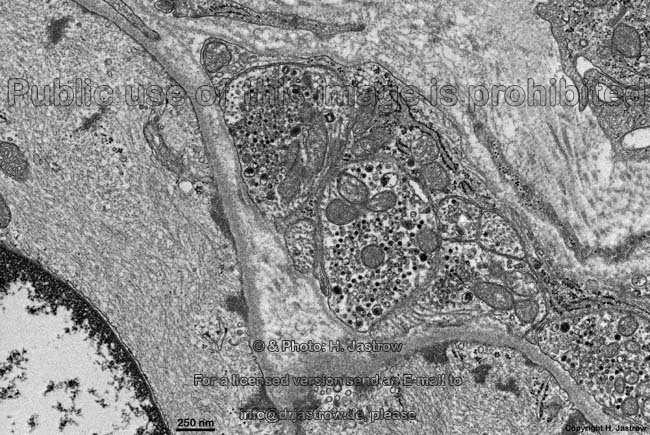
The endoneurium contains some fibrocytes
few fibroblasts, macrophages
and mast cells. The
collagen
fibrils in the endoneurium are usually oriented parallel to the nerve
fibres and are embedded in fundamental substance
which is poor in proteins and centrally reaches the cerebrospinal fluid
while in the periphery it opens into the connective
tissue which surrounds the nerve endings.
This is why it may carry e.g., herpes zoster viruses from spinal
ganglia into the dermatome of a nerve which then gets visible as shingles
(acute posterior ganglionitis). The endoneurium further may contain
capillaries,
few metarteriols and small
venules.
--> node of Ranvier, Schmidt-Lanterman
incisure, inner and outer
mesaxon
--> nerve tissue,
Schwann's
cells,
neuromuscular junction,
classification
of nerve fibres, CNS
--> Electron microscopic atlas Overview
--> Homepage of the workshop
Some images were kindly provided by Prof. H. Wartenberg;
other images, page & copyright H. Jastrow.