Overview veins (Venae):
(labelling in preparation)
 |
 |
 |
 |
 |
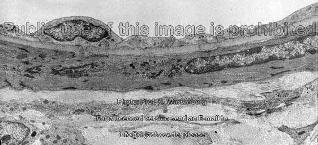
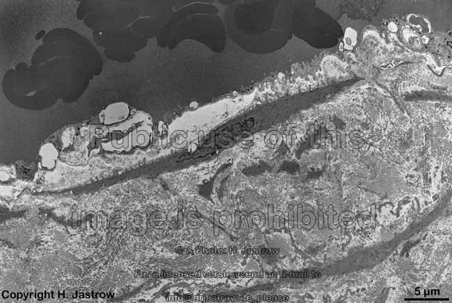
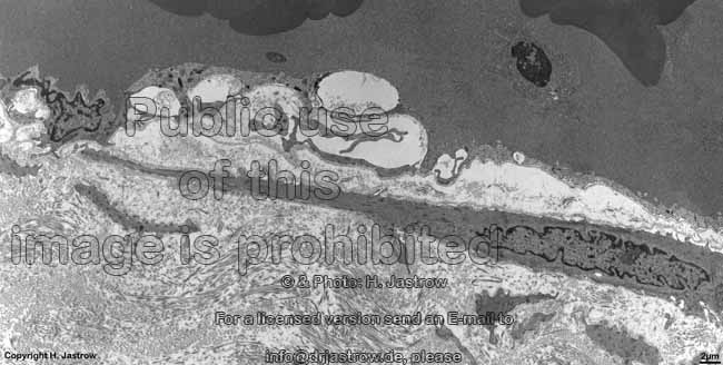
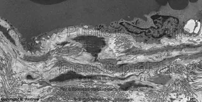
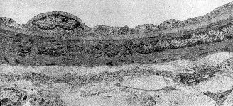
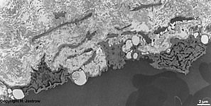
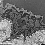
wall of a vein: endothelium + intima
and a long smooth muscle cell (monkey) |
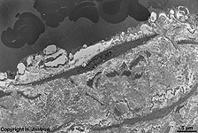
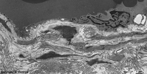
Intima of a human
vein at the ankle |
idem other area
(human) |
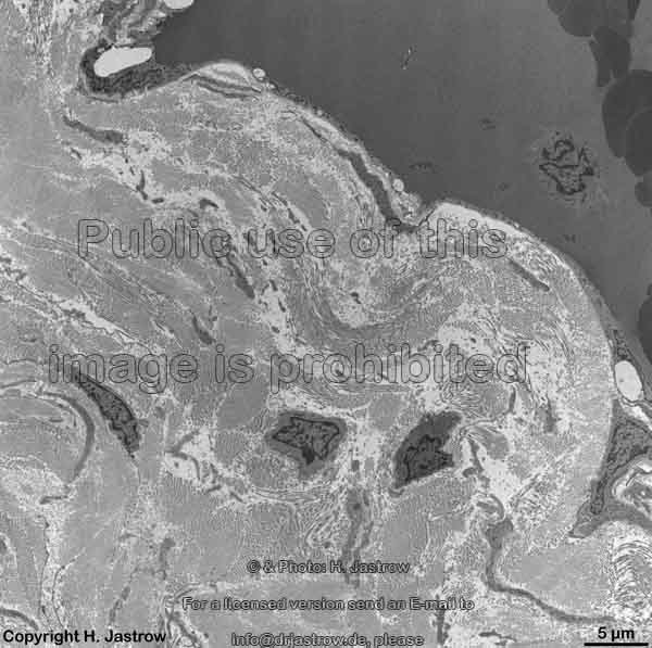
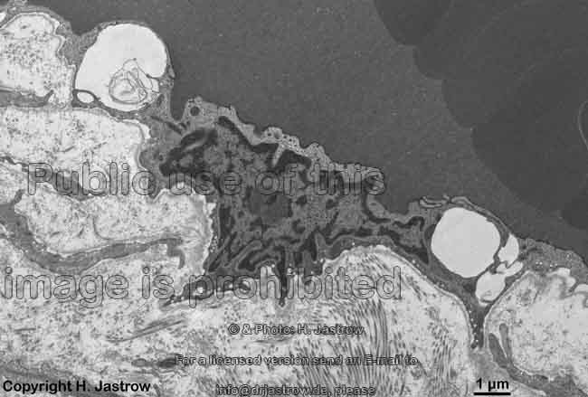
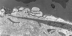
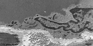
detail thereof: endothelial cells
showing sectioned nuclei (human) |
smooth muscle cell in the
subendothelial layer (human) |
 |
 |
 |
 |
 |
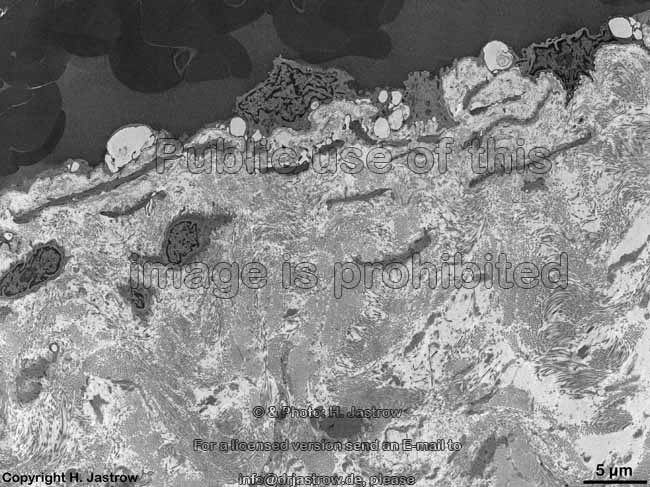
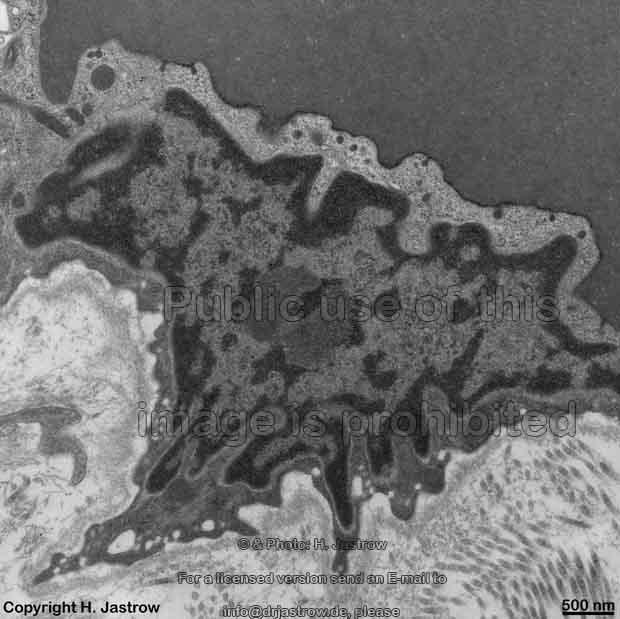
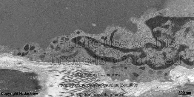
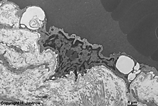
detail: smooth muscle cell + endothelial cells
with widened intercellular spaces (human) |
endothelial cell with widened
intercellular spaces (human) |
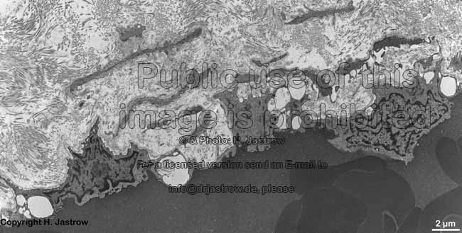
detail thereof:
nucleus |
subendothelial layer 2
(human) |
detail thereof: cytoplasm + organells
of the endothelial cell (human) |
Veins (Terminologia histologica: Venae) are large
blood vessels carrying blood
to the heart. With the exception
of the pulmonal veins which bring oxygenated blood to the left atrium of
the heart all other veins transport blood which is poor in oxygen. The
walls of veins resemble those of arteries, however since the blood pressure
is much lower in veins the different layers are thinner
and often less clearly bordered against each other.
Looking from the interior outwards the wall of a vein has the
following layers:
1. Tunica intima a squamous flatendothelium
borders the lumen. A
subendothelial layer (Stratum subendotheliale)
is located beyond its basal
lamina which is bordered by an internal elastic membrane (Membrana
elastica interna).
The Stratum subendotheliale (= Lamina propria intimae) has fine
reticularly ordered elastic fibres,
a lot of collagenous fibres and shows
singly lying intermingled smooth muscle cells.
In comparison to arteries the Membrana
elastica interna is quite poorly formed, often it is non-continuous,
and even may lack completely. The membrane consists of interconnected
elastic
fibres and marks the border to the media.
2. Tunica media consists of larger lamellas
and interconnected membranes of elastic fibres
and collagenous fibres many as well
as branched smooth muscle cells. In
comparison to arteries of equal diameter the media of veins is considerably
thinner and the amount of smooth muscle cells,
which are mainly in circular orientation, is reduced. The thickness of
this muscular layer is larger in veins which have to resist orthostatic
pressure (veins of the legs) and it is less in veins belonging to the gut.
3. Tunica externa (Adventitia) This
layer is strongest in veins of the abdominal cavity and otherwise also
remarkable. In contrast to arteries, there is no external elastic
membrane (Membrana elastica externa) on the border to the media
isolated fasciculi of elastic fibres are
present instead. In larger veins of the arm or legs or in the internal
jugulary vein elastic fibres form large
compact networks. The mainly loose connective tissue
has some amorphous fundamental substance several
larger and many smaller collagenous fibres
running in different directions. Further, fibrocytes
and free connective tissue cells are located
here. The adventitia serves for anchoring the veins to surrounding
tissues and contains small vessels for blood supply of the wall of the
vein itself (Vasa vasorum) as well as few non-myelinated nerve
fibres. Additional bundles of interconnected strands of longitudinally
oriented smooth muscle cells are encountered
in the adventitia of abdominal veins (V.
cava inferior,
V. portae,
V.
iliaca, V. renalis,
V.
lienalis) and V. azygos.
Most veins are of intermediate size,
i.e. have diameters of 2 - 9 mm. Such veins partly have a stronger
layer of elastic fibres (Membrana elastica
interna) with in some cases additional longitudinally oriented strands
of smooth muscle cells (Vv. popliteae,
femorales,
cephalicae,
mesenteriae, uterinae).
The circular to spirally oriented muscle fibre bundles of the media are
rich in cells in the veins of the leg whereas smooth
muscle cells are poor in veins of more upper regions of the body. However,
the adventitia is thick in most cases. There is considerable variation
in relative amount of elastic fibres, collagenous
fibres and intermingled smooth muscle cells.
Large veins with diameters of > 1 cm
usually show a thicker subendothelial layer of the intima with only few
smooth
muscle cells. Their adventitia is quite thick and in some cases shows
additional longitudinally oriented bundles of smooth
muscle cells. In case of the upper caval vein (V.
cava superior) a further outer layer of heart
muscle cells can be seen which increases in size towards the heart.
Beyond it longitudinally oriented bundles of smooth
muscle cells can be noted while in the media loose circularly oriented
smooth
muscle cells are present.
valves of veins are located in veins
of diameters from 3 to 8 mm to prevent a reflux of blood.
These valves are numerous in the extremities, especially in the foot region
since the orthostatic pressure (caused by the upright standing of humans)
is largest here. The valves are formed by half-moon shaped circular folds
of the intima including elastic and collagenous
fibres and are covered by an endothelium.
In case of heart insufficiency (weak heart) pressure in veins raises resulting
in an incomplete closing of the valves (insufficiency of venous valves).
This may lead to sac-like protrusions of the walls of veins which in most
cases are unilateral and which are called varices.
Veins with special wall construction:
- in the retina, the trabecules of the
spleen,
and in the Pia mater small veins
are seen which completely lack smooth muscle
cells. This is also the case in the venous sinuses of the
dura
(Sinus durae matris).
- in the network of veins running next to the spermatic chord (Plexus
pampiniformis), in the uterus (especially during pregnancy) and in the
umbilical vein an unusual large amount of smooth
muscle cells is present oriented circularly in the media and longitudinally
the adventitia.
- spiral strands of smooth
muscle cells of special strength which serve for (nearly) complete
obliteration of the vein called arresting veins are seen on the
base of the swell bodies. Under sexual arousal they serve for the swelling
of these bodies causing erection in males. Further, veins with similar
sphincter structures are present in the swell bodies of the nasal
mucosa, in veins of the adrenal gland medulla
and even in veins of the liver.
--> Venole, blood
vessels, endothelial cells
--> Electron microscopic atlas Overview
--> Homepage of the workshop
One image was kindly provided by Prof. H. Wartenberg;
other images, page & copyright H. Jastrow.