Tendinocyte of a tendon of a human eye muscle
(for unlabelled original image click here,
please!)
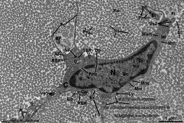
Af = Filamenta actinia (actin
filaments = intracellular microfilaments); C = Cytoplasma
(cytoplasm containing organells);
Clv = Vesiculum clathrinum (formation of a clathirn-coated
vesicle is visible in the region of this Ed);
Dp = Diaphragma pori (membrane of a nuclear
pore);
Ec = Euchromatinum (euchromatin;
merely electron-dense);
EF = Fibrilla elastica (elastic
fibril consists of central El = Elastin
[amorphous substance of elastic fibres]
which is surrounded
by individual fibrillin Fi microfilaments); Ex = Exocytoses
(exocytotic processes = transport
of vesicles out of a cell);
Fi = Fibrillin (extracellular microfibrils of connective
tissue);
G = Apparatus golgiensis (Golgi-apparatus);
Kf = collagen fibrils (of
type 1 and 3; note the different diameters of individual fibrils);
Ko = Fibrae collagenosae
(collagen fibres in cross-section; consist of bestehend aus collagen
fibrils which are interconnected
via proteoglycans Prg); Hc = Heterochromatinum (heterochromatin;
electron-dense); Ly = Lysosomes;
Mam = Matrix mitochondrialis (mitochondrial
matrix); Mi = Mitochondrion (mitochondrion
of intermediate type);
Mm = Membranae mitochondriales (mitochondrialmembranes;
the inner one has infoldings of variable widths into the Mam
further, there is an outer membrane); Mne = Membrana nuclearis
externa (outer nuclear membrane);
Mni = Membrana nuclearis interna (inner nuclear
membrane); N = Nucleus (nucleus);
P = Plasmalemma (cell membrane);
Pc
= Processus cellulares (immotile long thin processes of the tendinocyte
which contain Af and few organells); Pnu = Pori nucleares
(pores of the nuclear membrane; covered
with Dp);
Prg = proteoglykans (complexes of proteins with sugars which
glue Kf to each other); R = single ribosomes;
RER = rough endoplasmic reticulum
(intracellular network with bound ribosomes,
continues into SER near to G);
SER = smooth endoplasmic reticulum
(no ribosomes are attached here);
Sf = Substantia fundamentalis
(amorphous ground substance, contains mainly water and therefore is not
electron-dense);
Vs1 = Vesicula 1 (larger vesicles
with merely electron-dense content); Vs2 = Vesicula 2 (very small
vesicles).
The image shows a tendinocyte (Termonolgia histologica: Tendinocytus),
the characteristic cell of a tendon.
Tendinocytes are a kind of fibrocytes
that shows very long thin immotile processes (Pc) that diverge in
different directions between different collagen fibres (Ko). Tendinocytes
rest in place and have a low metabolic activity which is demonstated by
their poorness in cell organells. They synthetise the proteins which aggregate
outside the cells to form collagenous
and elastic fibrils. The latter are
rarely seen in tendons. These secreted basic proteins are so small that
they are not visible in the electron microscope, i.e. less than 0.5 nm
in diameter). Further tendinocytes secrete proteoglycans (Prg) and
other substances which form the amorphous ground substance (Substantia
fundamentalis Sfa). Some of these are stored in vesicles Vs
and transported to the cell membrane
(P) and then released by exocytosis
(Ex). Collagen fibres
(Ko) of tendons are oriented paralell to each other. Note that the
diameters of the fibrils vary considerably in diameter depending on the
amount of aggregated tropocollagen molecules of which they polymerise.
They are formed by interconnected collagen
fibrils (Kf) whereby proteoglycans (Prg) play a major
role in connections. Since the shown preparation is a cross-section no
longitudinally cut fibres are visible and thus the typical bands cannot
be seen here.
--> tendon
--> elastic - collagenous
fibres; ground substance; fibrocytes,
connective
tissue
--> Electron microscopic atlas Overview
--> Homepage of the workshop
Image, page & copyright H.
Jastrow.


