thoracic duct (rat)
(for unlabelled original image click here,
please!)
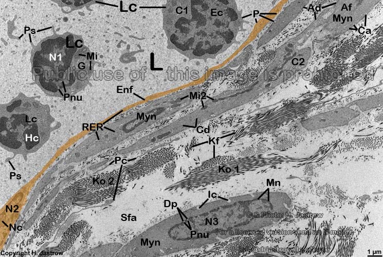
Ad = Areae densae (dense
bodies; anchoring Af at the cell
membrane);
Af = Filamenta actinia (actin
filaments in C2);
C1 = Cytoplasm (cellular
fluid + organelles of a lymphocyte); C2
= Cytoplasma (cytoplasm of a smooth
muscle cell);
Ca = Caveola (caveole;
small invagination of extracellular space
into the cytoplasm);
Cd = Corpuscula densa (fusiform
densities; intracellular interconnection of actin
filaments Af);
Dp = Diaphragma pori (membrane covering nuclear
pores);
Ec = Euchromatinum (euchromatin);
Enf = Endotheliocytus non fenestratus (non-fenestrated endothelial
cell);
G = Golgi-apparatus;
Hc
= Heterochromatinum (heterochromatin);
Ic = Invaginationes cellulae (larger invaginations of extracellular
space into a smooth muscle cell);
Kf = Fibrilla collageni (collagen
fibril);
Ko1 = Fibra collagenosa (collagen
fibre, tangential cut); Ko2 = Fibra collagenosa (collagen
fibre, cross-section)
L = Lumen ducti thoracici (lumen of the thoracic duct); Lc
= lymphocyti (lymphocytes);
Mi = Mitochondra (crista-type
of a lymphocyte); Mi2 = Mitochondra
(crista-type in smooth muscle cells);
Mn = Membrana nuclei (nuclear
membrane);
Myn = Myocyti non-striati (smooth
muscle cells);
N1 = nucleus
of a lymphocyte;
N2 = nucleus of an endothelial
cell; N3 = nucleus of a smooth
muscle cell; Nc = Nucleolus (Kernkörperchen);
P = Plasmalemmata (cell
membranes);
Pc = Processus cellulares (immobile processes of
fibrocytes);
Pnu = Pori nucleorum (nuclear
pores);
Ps = pseudopods
(mobile cell processes);
RER = rough endoplasmic reticulum
(intracellular meshwork of channels with attached ribosomes);
Sfa = Substantia fundamentalis amorpha (ground
substance).
The thoracic duct is the larges lymphatic vessel og the body. Its mane
comes from lat. Ductus = duct and Thorax = thorax. The duct collects lymph
from the intraabdominal organs, the pelvis and the legs. The thoracic duct
originates at the chymlic cistern (Cisterna chyli) and penetrates the diaphragm
to enter the thorax were it continues right to the aorta in posterior mediastinum.
The duct collects endothoracic lymph ducts as well. After a fat meal the
fluid in the duct becomes white (like milk). This is caused by thousands
of chylomicrones that originate from gut. The thoracic duct opens into
the left venous angle (Angulus venosus sinster) the junction of internal
jugular vein (V. jugularis interna und der V. subclavia sinistra. Thus
the whole content is directly delivered into the blood.
The thoracic duct has only a very thin monolayer of non-fenestrated
endothelial cells Enf and a
very weak media consisting of few
smooth
muscle cells Myn and larger amounts of
ground
substance Sfa. The media is attached to loose
connective tissue. There are only few corpuscules
in the lymph of the thoracic duct, in the present case exclusively
lymphocytes.
--> lymph vessels, blood vessels,
lymphocytes
--> Electron microscopic atlas Overview
--> Homepage of the workshop
Image, page & copyright H.
Jastrow.


